
Enzymatic Vs HPLC NAD+ Quantification For Research

Written By: Gary Hite, Research Content Writer
Reviewed By: Natalie Kunsman, M.D., Board-Certified Physician
Last Reviewed: August 5, 2026
Nicotinamide adenine dinucleotide (NAD+) plays a central role in cellular redox reactions, energy metabolism pathways, and enzymatic signaling processes that researchers study across multiple disciplines. Accurate quantification of NAD+ in biological samples is foundational to producing reliable, publishable data. Yet the choice between quantification methods is not always straightforward, and selecting the wrong approach for a given experimental context can introduce errors that compromise an entire study.
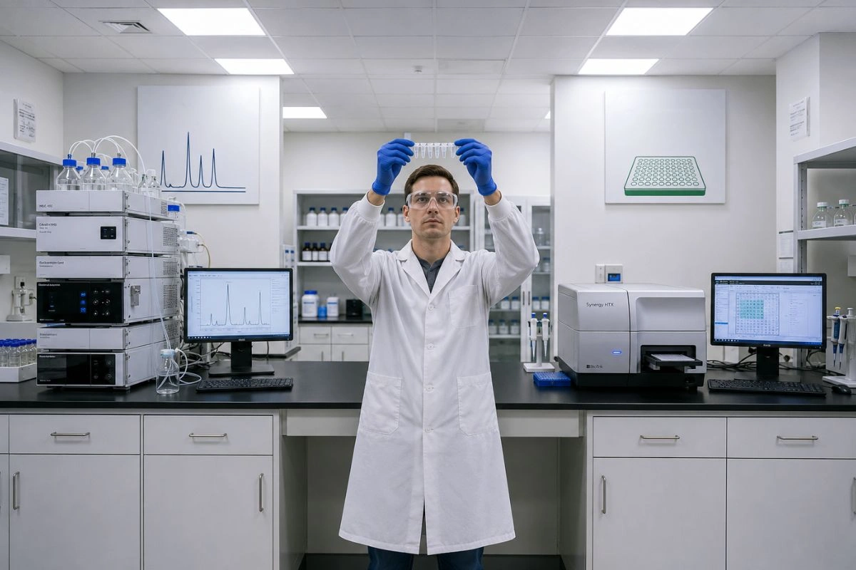
Two of the most widely used methods for NAD+ quantification in research settings are enzymatic cycling assays and high-performance liquid chromatography (HPLC). Each method offers distinct advantages, carries specific limitations, and presents its own set of common pitfalls that researchers must navigate.
This article provides a detailed comparison of these two approaches to help professional researchers and academic investigators make informed decisions when designing NAD+ quantification protocols.
Disclaimer: NAD+ is sold strictly for research purposes only. This compound is not intended for human consumption, therapeutic application, or diagnostic use. The following content is provided solely for educational and informational purposes directed at professional researchers and academic investigators. Nothing in this article constitutes medical advice or a claim of therapeutic benefit.
Enzymatic Cycling Assays: Principles and Performance
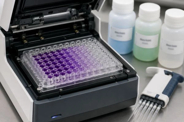
Enzymatic cycling assays for NAD+ quantification rely on a coupled reaction system in which NAD+ is continuously recycled between its oxidized and reduced forms. This cycling amplifies the signal proportionally to the NAD+ concentration in the sample, allowing detection at low concentrations. The reaction typically involves alcohol dehydrogenase or a similar enzyme paired with a colorimetric or fluorometric readout.
Sensitivity
Enzymatic assays are well regarded in the research literature for their sensitivity. The cycling amplification mechanism allows detection of NAD+ at picomolar to low nanomolar concentrations, depending on the specific assay configuration and detection method. Fluorometric detection generally achieves lower limits of detection than colorimetric approaches. For researchers working with limited sample volumes or low-abundance biological matrices, enzymatic assays often provide the sensitivity needed to generate quantitative data.
Throughput
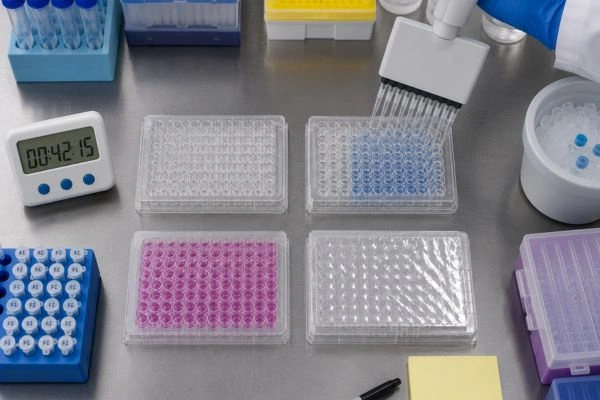
One of the practical advantages of enzymatic cycling assays is their compatibility with microplate formats. Researchers can process 96 or 384 samples simultaneously using a standard plate reader, making this approach suitable for experiments that require high throughput. Sample preparation is generally less complex than chromatographic methods, and total assay time from sample addition to data acquisition can be as short as 30 to 60 minutes under optimized conditions.
Common Pitfalls
Despite their advantages, enzymatic assays carry specific risks that researchers should actively manage. Interference from NADH, NADP+, and NADPH can inflate or distort NAD+ readings if the assay does not adequately discriminate between these closely related metabolites. Sample matrix effects, including pH shifts, protein content, and the presence of enzyme inhibitors, can alter cycling efficiency and skew results. Researchers should validate each new sample type with spike-and-recovery experiments to confirm that the matrix does not compromise assay performance.
Additionally, lot-to-lot variability in commercial enzyme reagents can affect reproducibility between experiments. Documenting reagent lot numbers and running fresh standard curves with every plate are essential practices for maintaining data quality.
HPLC-Based Quantification: Principles and Performance
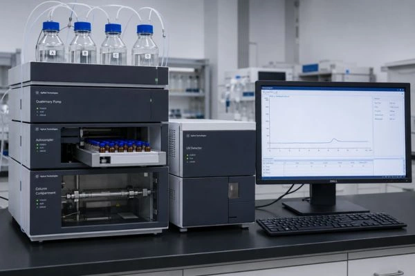
HPLC-based NAD+ quantification separates the target analyte from other sample components using a chromatographic column before detection, typically by UV absorbance at 254 or 260 nm. Reverse-phase and ion-pairing chromatographic methods are the most common configurations for NAD+ analysis in research literature.
Sensitivity
HPLC methods generally offer detection limits in the low nanomolar to micromolar range for NAD+, depending on the column chemistry, mobile phase composition, injection volume, and detector type. While standard UV detection is adequate for many research applications, coupling HPLC with mass spectrometry (LC-MS) can push sensitivity significantly lower, reaching picomolar levels comparable to enzymatic assays. However, LC-MS adds cost, complexity, and maintenance requirements that may not be justified for every laboratory or research question.
Throughput
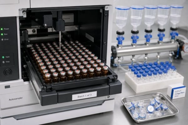
Throughput is one of the primary tradeoffs with HPLC-based methods. Individual chromatographic runs for NAD+ typically range from 10 to 30 minutes per sample, depending on the method and column conditions. Even with autosamplers, processing a full sample set of 50 to 100 samples can require an entire day of instrument time. Sample preparation for HPLC is also more involved, often requiring protein precipitation, filtration, and careful pH adjustment before injection. For research programs that prioritize throughput and rapid turnaround, this time investment is a meaningful consideration.
Common Pitfalls
HPLC-based NAD+ quantification introduces its own set of challenges. Co-elution of NAD+ with structurally similar metabolites (such as NADH or ADP-ribose) can lead to inaccurate quantification if the chromatographic separation is not fully optimized. Researchers should validate peak identity using spiked standards and, where possible, confirm analyte identity with a secondary detection method such as mass spectrometry.
Column degradation over time can shift retention times and reduce resolution, leading to gradual loss of method performance. Maintaining a column logbook and replacing columns on a defined schedule based on injection count or performance metrics helps prevent this issue from going undetected.
Mobile phase preparation errors, including incorrect buffer concentrations or pH values, can dramatically alter elution profiles. Preparing mobile phases fresh from validated protocols and verifying pH before each run are simple but critical quality control steps.

Choosing the Right Method for Your Research Design
Neither enzymatic assays nor HPLC is universally superior for NAD+ quantification. The right choice depends on the specific demands of each research project. Consider the following decision factors when selecting your approach.
If your primary need is high throughput with moderate to high sensitivity, and your sample matrix is well characterized, enzymatic cycling assays are typically the more efficient option. They require less specialized equipment, integrate easily with plate-based workflows, and deliver faster time to data.
If your research requires definitive analyte identification, separation from closely related metabolites, or quantification of multiple NAD-related species within the same run, HPLC-based methods provide the chromatographic resolution needed to meet those requirements. The additional investment in time and instrument access is justified when specificity and multi-analyte capability are priorities.
For the highest confidence in NAD+ quantification, some research groups employ both methods in parallel, using enzymatic assays for primary screening and HPLC for confirmation of key findings. This orthogonal validation strategy strengthens the reliability of published data and helps identify discrepancies early in the experimental pipeline.
Practical Recommendations for Reproducible NAD+ Quantification
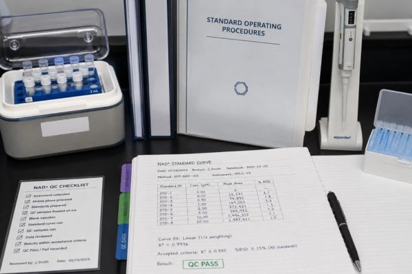
Regardless of the method selected, the following practices support data integrity in NAD+ research. Always run standard curves with every assay session or HPLC batch. Include matrix-matched quality control samples at known concentrations. Document all reagent sources, lot numbers, instrument settings, and sample preparation steps. Perform method validation, including linearity, precision, accuracy, and limit of detection, before applying a new assay to experimental samples. Store all samples under conditions that minimize NAD+ degradation, typically flash-frozen and kept at minus eighty degrees Celsius until analysis.
Commit to these standards, and your NAD+ quantification data will stand up to the scrutiny that rigorous peer review demands.
Conclusion
Choosing between enzymatic cycling assays and HPLC for NAD+ quantification is not a matter of preference. It is a methodological decision that shapes the quality, specificity, and reproducibility of every data point your study produces. Match your method to your research requirements. Use enzymatic assays when throughput and sensitivity in well-characterized matrices are priorities. Use HPLC when analyte specificity, metabolite separation, or multi-analyte profiling drives your experimental design. When the stakes demand maximum confidence, deploy both methods in parallel to cross-validate key findings. Regardless of which approach you select, run fresh standard curves with every session, include matrix-matched quality controls, validate method performance before applying it to experimental samples, and document every variable from reagent lot numbers to mobile phase pH. NAD+ quantification is only as reliable as the protocols surrounding it. Build those protocols with precision, enforce them without exception, and let reproducible data define your research output.
Frequently Asked Questions
When should I choose an enzymatic cycling assay over HPLC for NAD+ quantification?
Choose enzymatic assays when your research requires high throughput, your sample matrix is well characterized, and you need sensitivity in the picomolar to low nanomolar range. Enzymatic assays integrate with 96- or 384-well microplate formats, process large sample sets in 30 to 60 minutes, and require less specialized equipment than chromatographic methods. However, validate each new sample type with spike-and-recovery experiments first to confirm that matrix effects do not compromise cycling efficiency or distort your readings.
What are the main advantages of HPLC for NAD+ research compared to enzymatic methods?
HPLC provides chromatographic separation that distinguishes NAD+ from structurally similar metabolites such as NADH, NADP+, and ADP-ribose within the same analytical run. This specificity is critical when your research question requires definitive analyte identification or simultaneous quantification of multiple NAD-related species. Coupling HPLC with mass spectrometry further extends sensitivity into the picomolar range while adding a secondary layer of analyte confirmation that enzymatic assays cannot provide on their own.
How do I prevent interference from related metabolites in enzymatic NAD+ assays?
Interference from NADH, NADP+, and NADPH is one of the most common pitfalls in enzymatic quantification. Select assay kits or reaction configurations that include specific extraction or decomposition steps to eliminate these interfering species before the cycling reaction begins. Run spike-and-recovery experiments with each related metabolite at physiologically relevant concentrations to quantify cross-reactivity. If interference exceeds acceptable thresholds, consider switching to HPLC or adding a sample cleanup step before the enzymatic assay.
Can I use both enzymatic and HPLC methods together in the same study?
Yes, and many research groups do exactly this for orthogonal validation. Use enzymatic cycling assays as your primary screening tool to process large sample sets efficiently, then confirm critical findings with HPLC to verify analyte identity and rule out interference. This parallel approach strengthens data reliability, catches discrepancies early in the experimental pipeline, and produces results that hold up under rigorous peer review. Document both methods fully so reviewers and other investigators can assess and reproduce your validation strategy.
What storage and handling practices protect NAD+ sample integrity before quantification?
Flash-freeze all biological samples immediately after collection and store them at minus eighty degrees Celsius until analysis. NAD+ degrades rapidly at elevated temperatures, at neutral to alkaline pH, and through repeated freeze-thaw cycles. Aliquot samples into single-use volumes at the time of collection to avoid repeated freezing and thawing. Record the time between sample collection and freezing, the number of freeze-thaw events, and storage duration for every sample. These variables directly affect measured NAD+ concentrations and must be controlled and documented to ensure data integrity across your study.