❌ Not for Human Consumption
●
✅ For Research Use Only
●
All products are intended strictly for laboratory research purposes only
●
❌ Not for Human Consumption
●
✅ For Research Use Only
●
All products are intended strictly for laboratory research purposes only
●

Analytical Characterization Of Recombinant HGH: SDS-PAGE, RP-HPLC, And Mass Spectrometry Methods

Written By: Gary Hite, Research Content Writer
Reviewed By: Natalie Kunsman, M.D., Board-Certified Physician
Last Reviewed: September 17, 2026
Research Use Only. All products and information referenced in this article are intended strictly for in vitro laboratory research and educational purposes. The recombinant HGH discussed here is supplied and described as a research reagent only. It is not a drug, food, dietary supplement, or cosmetic, and it is not intended for human consumption, veterinary use, diagnostic procedures, or any clinical application. This content makes no medical claims, describes no therapeutic uses, and is not intended to diagnose, treat, cure, or prevent any condition. Handling is restricted to qualified professional researchers and academic personnel operating in a controlled laboratory environment.
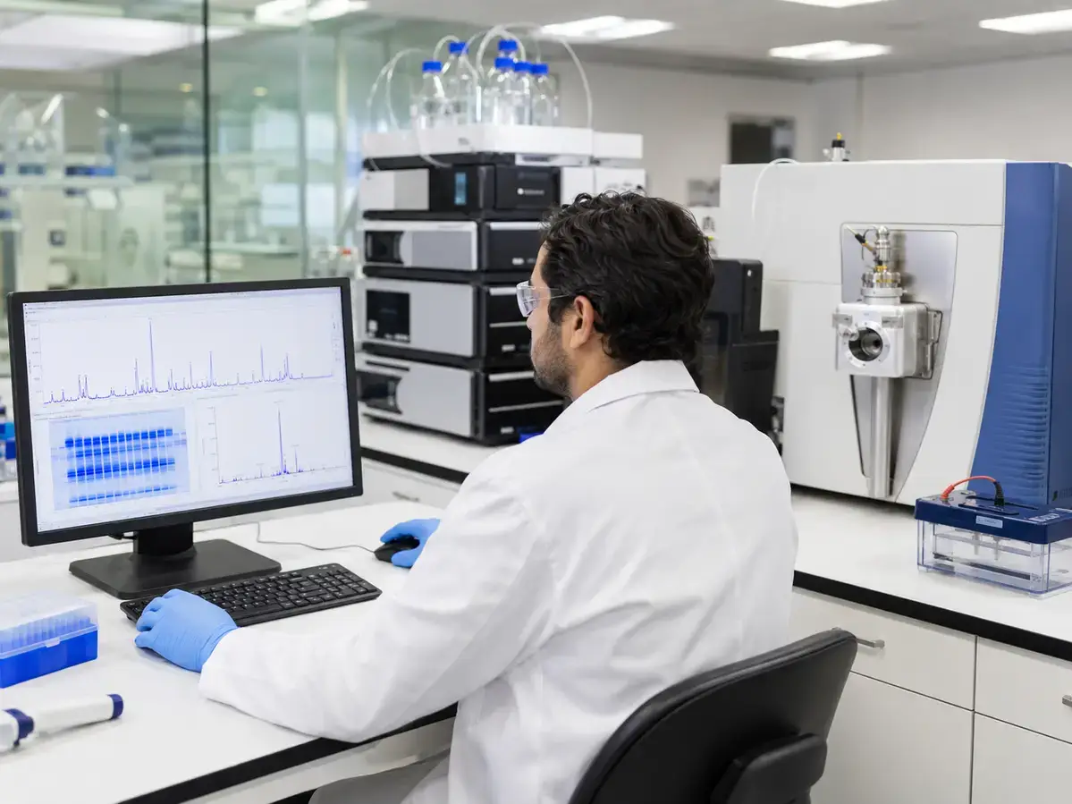
Recombinant human growth hormone (recombinant HGH) is one of the most extensively studied recombinant proteins in the life sciences, which makes it a frequent reference analyte in protein chemistry laboratories. For research teams that handle this 191 residue polypeptide as an experimental reagent or as a characterization standard, analytical rigor is the foundation of reproducible work. Before any downstream experiment, researchers need to confirm three things: that the material is what the label says it is, that it is sufficiently pure, and that its higher order structure is intact.
This article walks through three complementary analytical techniques that laboratories rely on to answer those questions: SDS-PAGE, reversed phase high performance liquid chromatography (RP-HPLC), and mass spectrometry. Use this guide to plan a characterization workflow, interpret common results, and understand why no single method tells the whole story. Everything that follows is written for analytical chemists, protein scientists, and academic researchers working strictly within a research use context.
Understanding the Analyte Before You Run a Single Assay
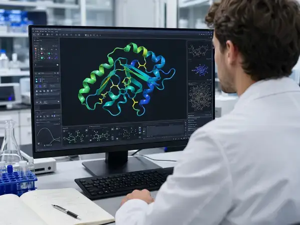
Strong characterization starts with knowing the molecule. Recombinant HGH is a single chain, non glycosylated polypeptide of 191 amino acids with a theoretical molecular mass of roughly 22,124 Da, which is why the protein is often referred to as the 22 kDa form. It carries two intramolecular disulfide bonds that stabilize its folded structure, and a smaller 20 kDa isoform produced by alternative splicing is well documented in the literature. Knowing these baseline properties lets researchers set expectations for every method below.
Map out the quality attributes you intend to monitor before selecting your methods. For recombinant protein reference material, research teams commonly track apparent molecular weight, monomer purity, aggregation and fragmentation, charge or sequence variants, and chemical modifications such as methionine oxidation, asparagine or glutamine deamidation, and N terminal heterogeneity. Each technique in this article addresses a different subset of those attributes, so define your questions first and then choose the tools that answer them.
SDS-PAGE: Establishing Apparent Molecular Weight and Purity
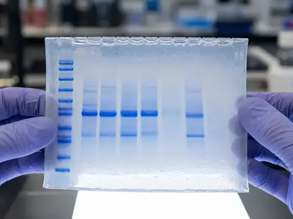
Sodium dodecyl sulfate polyacrylamide gel electrophoresis (SDS-PAGE) is usually the first orthogonal check a laboratory performs because it is fast, accessible, and highly informative. The method coats the protein in negatively charged SDS, which masks intrinsic charge and drives separation primarily by molecular size as the sample migrates through the polyacrylamide matrix under an electric field.
How to Set Up the Separation
Select a gel percentage suited to a 22 kDa target. Many laboratories run a 12 to 15 percent resolving gel, or a gradient gel, to resolve the recombinant HGH band cleanly from lower mass fragments and higher mass dimers. Always run a molecular weight ladder in an adjacent lane so you can estimate apparent mass and confirm that the principal band migrates near the expected position.
Run samples under both reducing and non reducing conditions to extract more structural information. Under reducing conditions, a reagent such as dithiothreitol or beta mercaptoethanol breaks the disulfide bonds, which can shift migration and reveal covalently linked species. Comparing the two conditions helps researchers distinguish disulfide linked dimers from non covalent aggregates and gives a clearer picture of covalent integrity.
What the Gel Tells You
Read the gel for both identity and purity signals. A single, sharp, dominant band at the expected apparent mass supports identity, while faint additional bands point to fragments, dimers, or process related species. To quantify purity, scan the stained gel and apply densitometry, which expresses the main band as a percentage of total stained protein in the lane. Choose your stain based on sensitivity needs: Coomassie is robust and quantitative for routine work, while silver staining offers far greater sensitivity when you need to detect low level impurities.
Interpret SDS-PAGE results with its limitations in mind. Apparent molecular weight from a gel is an estimate rather than an exact mass, because SDS binding and migration behavior can differ between proteins. Treat the gel as a powerful screening and purity tool, and confirm exact mass with the orthogonal methods that follow.
RP-HPLC: Resolving Purity and Related Substances
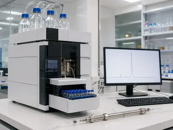
Reversed phase high performance liquid chromatography (RP-HPLC) separates molecules by hydrophobicity, which gives it resolving power that complements the size based separation of SDS-PAGE. Because subtle modifications can change a protein’s surface chemistry, RP-HPLC often resolves variants that co migrate on a gel, making it a workhorse for purity and related substances analysis.
Building a Reliable Method
Choose a column chemistry matched to the analyte. Wide pore C4 columns are a common starting point for intact proteins and larger polypeptides, while C18 chemistries can offer additional retention for certain applications. Pair the column with a mobile phase system built on water and acetonitrile, modified with an ion pairing acid such as trifluoroacetic acid or formic acid to sharpen peak shape and improve reproducibility.
Develop a gradient that elutes the main peak with good resolution from neighboring species, and set a column temperature and flow rate that keep retention times stable across a sequence. Monitor the eluate by UV detection at 214 nm, which responds to the peptide backbone, and at 280 nm, which responds to aromatic residues. Running both wavelengths gives a fuller view of the sample and aids peak assignment.
Interpreting the Chromatogram
Evaluate the main peak and everything around it. Report main peak purity as a percentage of total peak area, and characterize the related substances that elute before and after it, since these can correspond to oxidized, deamidated, or otherwise modified forms. Use a well characterized reference standard run under identical conditions to support identity by retention time, and confirm method performance with system suitability checks before you trust the data.
Treat method robustness as part of the result. Small shifts in mobile phase composition, temperature, or column age can move retention times, so document conditions carefully and verify reproducibility across replicate injections. Consistent, well controlled RP-HPLC data is one of the strongest purity readouts available to a protein laboratory.
Mass Spectrometry: Confirming Identity at the Molecular Level
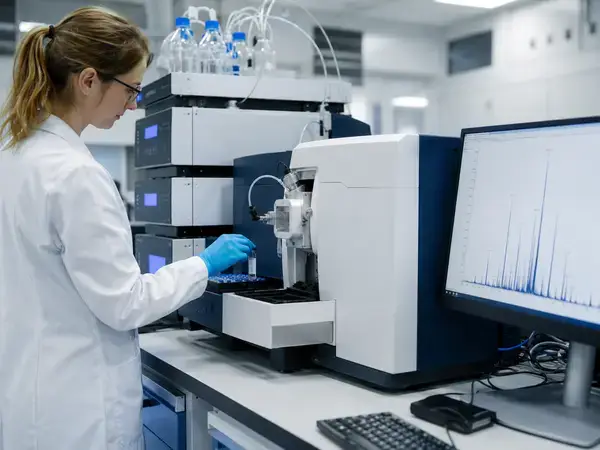
Mass spectrometry (MS) provides the most direct confirmation of molecular identity because it measures mass with high accuracy. Where SDS-PAGE estimates size and RP-HPLC reports relative purity, MS pins down the exact mass and, with the right workflow, the underlying sequence and modifications.
Intact Mass Analysis
Start with intact mass measurement to confirm the molecular weight of the whole protein. Electrospray ionization (ESI) generates a series of multiply charged ions that software deconvolutes into a single average or monoisotopic mass, which researchers compare against the theoretical mass near 22,124 Da for the 22 kDa form. Matrix assisted laser desorption ionization time of flight (MALDI-TOF) offers an alternative intact mass approach that tolerates certain sample conditions well. A measured intact mass that matches the expected value is strong evidence of identity and can flag mass shifts caused by modifications.
Peptide Mapping and Sequence Confirmation
Move to peptide mapping when you need sequence level confidence. In a bottom up workflow, an enzyme such as trypsin cleaves the protein into defined peptides, which are then separated and analyzed by liquid chromatography tandem mass spectrometry (LC-MS/MS). Matching the observed peptides to the expected sequence builds sequence coverage and confirms primary structure with high specificity.
Use the same workflow to localize and quantify modifications. Peptide mapping can pinpoint sites of methionine oxidation, asparagine deamidation, and N terminal variation, and dedicated experiments can map disulfide bond connectivity. High resolution accurate mass instruments such as Orbitrap and quadrupole time of flight platforms make these assignments more confident by resolving closely spaced species. Together, intact mass and peptide mapping give a layered picture: the whole molecule plus the fine detail.
Building an Orthogonal Characterization Workflow
No single technique delivers a complete characterization, which is the central reason laboratories combine all three. SDS-PAGE provides a fast purity and size screen, RP-HPLC resolves variants and quantifies related substances by hydrophobicity, and mass spectrometry confirms exact mass and sequence. Because each method separates or measures on a different principle, agreement across all three gives far greater confidence than any one result alone.
Sequence your workflow to build evidence efficiently. Many teams begin with SDS-PAGE for a rapid integrity and purity check, advance to RP-HPLC for quantitative purity and variant resolution, and reserve mass spectrometry for definitive identity and structural confirmation. Anchor the entire workflow to a well characterized reference standard, run appropriate controls, and capture system suitability data so results are comparable across analysts, instruments, and days.
Document everything in a way that supports reproducibility. Record sample handling, buffer composition, instrument parameters, and acceptance criteria, and retain raw data alongside processed results. Disciplined documentation turns a set of individual assays into a defensible characterization package that other researchers can understand and repeat.
Best Practices for Reproducible Results
Apply a few core practices to keep data clean and comparable. Standardize sample preparation and storage to limit handling artifacts, since freeze thaw cycles and inconsistent buffers can introduce aggregation or modification that confound your readouts. Run replicates rather than single measurements, include reference standards on every plate or sequence, and validate or qualify your methods before relying on them for comparative work.
Calibrate expectations to each method’s strengths. Use SDS-PAGE for accessible screening, RP-HPLC for high resolution purity, and mass spectrometry for definitive identity, and let the three datasets cross check one another. When results disagree, treat the conflict as useful information that points you toward sample heterogeneity, a method artifact, or a parameter that needs tightening.
Conclusion
Effective analytical characterization of recombinant HGH depends on combining complementary techniques rather than trusting any single result. Run SDS-PAGE first to screen apparent molecular weight, monomer purity, and covalent integrity, then apply RP-HPLC to resolve related substances and quantify main peak purity by hydrophobicity. Confirm identity at the molecular level with mass spectrometry, using intact mass analysis to verify the expected mass near 22,124 Da and peptide mapping to validate sequence and localize modifications. Anchor every step to a well characterized reference standard, document your conditions in full, and run replicates so results stay reproducible across analysts and instruments. When the three datasets agree, you gain confidence that your material is correctly identified, sufficiently pure, and structurally intact. Treat this orthogonal workflow as the backbone of disciplined protein characterization, and remember that all work described here is intended strictly for laboratory research and educational purposes, never for human consumption.
FAQs
What are the main methods for analytical characterization of recombinant HGH?
Researchers rely on three complementary techniques. Run SDS-PAGE to screen apparent molecular weight and purity, use RP-HPLC to resolve related substances and quantify main peak purity, and apply mass spectrometry to confirm exact mass and sequence. Combining all three delivers far stronger characterization than any single assay.
What is the expected molecular weight of recombinant HGH?
The standard form is a single chain, non glycosylated polypeptide of 191 amino acids with a theoretical average mass of approximately 22,124 Da, commonly described as the 22 kDa form. Confirm this value with intact mass spectrometry by comparing the measured mass against the theoretical mass.
How do researchers determine the purity of recombinant HGH?
Use orthogonal readouts. RP-HPLC reports main peak purity as a percentage of total peak area, while densitometry of an SDS-PAGE gel expresses the main band as a percentage of total stained protein. Run both and cross check the results to build a reliable purity picture.
Why use multiple orthogonal techniques instead of a single method?
Each method measures a different property, so no single technique tells the whole story. SDS-PAGE estimates size, RP-HPLC separates by hydrophobicity, and mass spectrometry confirms exact mass and sequence. When all three datasets agree, you gain high confidence that the material is correctly identified, sufficiently pure, and structurally intact.
Is recombinant HGH intended for any use beyond research?
No. The recombinant HGH described here is supplied strictly as a research reagent for in vitro laboratory and educational purposes only. It is not for human consumption, veterinary use, diagnostic procedures, or any clinical application, and this content makes no medical claims.