❌ Not for Human Consumption
●
✅ For Research Use Only
●
All products are intended strictly for laboratory research purposes only
●
❌ Not for Human Consumption
●
✅ For Research Use Only
●
All products are intended strictly for laboratory research purposes only
●

Mass Spectrometry Characterization Of Hexarelin Acetate

Written By: Gary Hite, Research Content Writer
Reviewed By: Natalie Kunsman, M.D., Board-Certified Physician
Last Reviewed: September 25, 2026
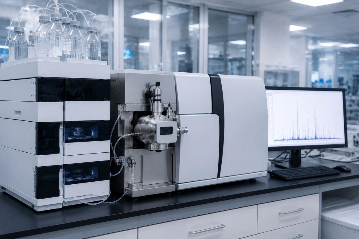
Mass spectrometry has become an indispensable tool for confirming the identity and structure of synthetic peptides, and for laboratories working with Hexarelin Acetate it provides a level of molecular confidence that few other techniques can match. Where chromatography measures how pure a sample is, mass spectrometry answers a different question: is this the correct molecule, and what exactly is present alongside it. This article explains how mass spectrometry characterization is applied to Hexarelin Acetate, a synthetic research peptide supplied as a lyophilized powder, covering ionization techniques, mass analyzer selection, molecular weight confirmation, sequence analysis by tandem mass spectrometry, and the interpretation of results in a controlled laboratory setting.
Disclaimer: Hexarelin Acetate is supplied and intended strictly for in vitro laboratory research and educational purposes only. It is not a drug, food, dietary supplement, or cosmetic, and it is not intended for human or veterinary use, consumption, diagnostic application, or therapeutic application. This material is intended for handling exclusively by qualified researchers and academic professionals within a controlled laboratory setting. The information below is educational and addresses laboratory analysis only. It does not constitute medical, clinical, or professional advice.

Understanding Hexarelin Acetate as a Research Material
Hexarelin is a synthetic hexapeptide investigated in laboratory research related to growth hormone secretagogue receptor signaling. It is typically supplied as the acetate salt in lyophilized form, with the peptide portion carrying a molecular weight of approximately 887 Daltons. Several of its structural features are directly relevant to mass spectrometry. The sequence contains aromatic residues, including tryptophan and phenylalanine, and it terminates in a C-terminal amide rather than a free carboxylic acid. These details define the exact mass a researcher expects to observe and shape the fragmentation behavior seen during analysis. Knowing the molecular characteristics of the material is the foundation for designing and interpreting any characterization workflow.
Why Mass Spectrometry Is Central to Peptide Characterization
Chromatographic methods establish how much of a sample is the intended compound, but they do not by themselves confirm what that compound is. Mass spectrometry measures the mass-to-charge ratio of ionized molecules, allowing a researcher to determine molecular weight with high precision and to compare the observed value against the theoretical mass of the target peptide. This makes it the primary technique for identity confirmation.
Beyond confirming molecular weight, mass spectrometry detects and helps characterize impurities and adducts, and through fragmentation it can verify the amino acid sequence itself. Used alongside chromatography, it completes the analytical picture: chromatography reports purity, and mass spectrometry confirms identity and structure. Together they form the core of rigorous peptide characterization.
Ionization Techniques for Peptide Analysis
A molecule must be converted into a gas-phase ion before it can be measured. For peptides, soft ionization techniques are preferred because they transfer the molecule into the gas phase while keeping it intact rather than breaking it apart. Two methods dominate peptide work.
Electrospray Ionization (ESI)
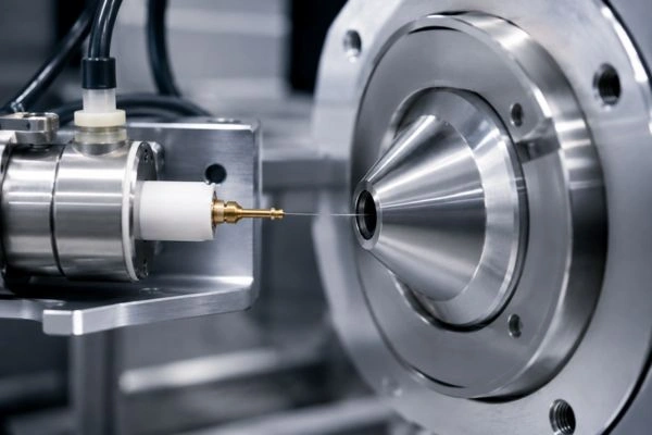
Electrospray ionization generates ions by applying a high voltage to a fine spray of sample solution, producing charged droplets that release ions as the solvent evaporates. ESI is a soft technique that frequently produces multiply charged ions, which is advantageous because it brings larger molecules into the measurable mass-to-charge range. A defining strength of ESI is its compatibility with liquid chromatography, enabling coupled LC-MS workflows in which a sample is separated and then analyzed in a single continuous process.
Matrix-Assisted Laser Desorption/Ionization (MALDI)
Matrix-assisted laser desorption ionization mixes the sample with a light-absorbing matrix compound, then uses a laser pulse to desorb and ionize the molecules. MALDI is also a soft technique and typically produces singly charged ions, which can simplify spectral interpretation. It is commonly paired with a time-of-flight analyzer and is well suited to rapid molecular weight confirmation of peptides. Selecting between ESI and MALDI depends on the analytical goal, the available instrumentation, and whether online separation is required.

Selecting a Mass Analyzer
The mass analyzer determines how ions are separated and measured, and the choice influences resolution, mass accuracy, and the type of information obtained. Several analyzer types are common in peptide characterization:
- Time-of-flight (TOF) analyzers measure the time ions take to travel a fixed distance, offering high resolution and good mass accuracy. They are frequently paired with MALDI.
- Quadrupole analyzers act as tunable mass filters and are robust and widely used, particularly within triple quadrupole instruments configured for targeted analysis.
- High-resolution analyzers such as Orbitrap and Fourier transform systems deliver high-resolution accurate mass measurements that support confident molecular formula confirmation.
- Hybrid instruments, including quadrupole time-of-flight and quadrupole Orbitrap configurations, combine filtering and high-resolution detection to support both identity confirmation and detailed structural work.
For confirming the identity of a defined peptide, a high-resolution instrument provides the mass accuracy needed to distinguish the target from closely related species.
Confirming Molecular Weight and Identity
Begin identity confirmation by comparing the measured mass to the calculated mass of the peptide. Two values are relevant here. The average mass is calculated using the natural isotopic abundances of the constituent elements, while the monoisotopic mass uses the most abundant isotope of each element. High-resolution instruments typically report the monoisotopic mass, which is calculated directly from the molecular formula and provides a precise target for comparison.
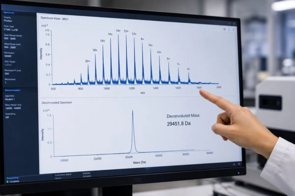
When ESI produces multiply charged ions, the spectrum will show a series of peaks corresponding to different charge states, such as singly and doubly protonated forms. Deconvolution software converts this series into a single neutral mass, which is then compared to the theoretical value. Mass accuracy is expressed in parts per million, and high-resolution instruments can achieve agreement within a few parts per million. A measured mass that matches the calculated mass within the accepted tolerance provides strong confirmation that the analyzed material is the intended peptide. Confirming the expected C-terminal amide, which shifts the mass relative to a free-acid form, is one example of how a structural feature is verified through accurate mass measurement.

Sequence Confirmation with Tandem Mass Spectrometry
Accurate mass confirms what a molecule weighs, but tandem mass spectrometry, often written as MS/MS, confirms how it is assembled. In this approach, a selected ion is fragmented inside the instrument, commonly through collision-induced dissociation or related techniques, and the resulting fragment ions are measured.
Peptide backbones fragment in predictable ways, producing ion series that correspond to cleavage at the peptide bonds. The fragments retaining the N-terminus and those retaining the C-terminus form complementary series that, when mapped, reconstruct the amino acid sequence and confirm it against the expected structure. This is particularly useful for a peptide such as Hexarelin Acetate, which includes modified residues and a C-terminal amide whose presence should be reflected in the fragmentation pattern.
One important limitation deserves mention. Mass spectrometry measures mass, so it cannot distinguish stereoisomers that share an identical molecular formula. Determining the configuration of D- and L-amino acids requires orthogonal techniques rather than mass measurement alone. Recognizing this boundary is part of interpreting results responsibly.

Sample Preparation for Mass Spectrometry
Prepare samples with the ionization method in mind. For electrospray ionization, dissolve the material in volatile, mass-spectrometry-compatible solvents and avoid non-volatile salts and buffers, which suppress signal and contaminate the source. Where a sample contains residual ion-pairing agents from earlier processing, desalting or substituting a more compatible modifier such as formic acid can substantially improve signal quality. Sample concentration should suit the instrument and the analysis, and gentle handling helps avoid introducing degradation that could be misread as an impurity. When characterization is performed by LC-MS, the chromatographic step provides online separation immediately ahead of detection, which simplifies the analysis of samples that contain multiple components.
Identifying Impurities and Adducts
Mass spectrometry is well suited to recognizing species that accompany the main peptide, because each appears at a characteristic mass difference. Adducts formed with sodium or potassium shift the observed mass upward by predictable amounts relative to the protonated form. Oxidation adds a defined mass increment associated with the addition of oxygen, often at susceptible residues such as tryptophan. Deamidation produces a small mass increase, and process-related deletion sequences appear as mass differences corresponding to the missing residues. By interpreting these differences, a researcher can distinguish true impurities from instrument artifacts and build a clearer understanding of sample composition than purity data alone would allow.

Interpreting and Documenting Results
Sound interpretation rests on a clear comparison between observed and theoretical values within a defined mass tolerance, supported wherever possible by a well-characterized reference standard. Record the instrument type, ionization method, mass analyzer, acquisition parameters, and the software used for deconvolution and interpretation. Document both the confirmed molecular mass and, where performed, the sequence-confirming fragment data. A certificate of analysis for a research material commonly reports identity confirmation by mass spectrometry, and researchers should treat it as a baseline while confirming results under their own conditions when their work requires it. Thorough documentation ties each conclusion back to a defined, repeatable procedure and supports reproducibility across a research program.
Conclusion
Mass spectrometry gives peptide researchers a direct, high-confidence route to confirming that a material is what it is intended to be. Choose an ionization technique suited to the workflow, select a mass analyzer matched to the required resolution, confirm molecular weight by comparing observed and theoretical masses, and use tandem mass spectrometry to verify sequence where structural detail is needed. Prepare samples for clean ionization, interpret impurities and adducts by their characteristic mass differences, and document every parameter. Applied with discipline, and paired with chromatographic purity data, these methods deliver the molecular certainty that rigorous research depends on.
Disclaimer: This content is provided for educational and informational purposes regarding laboratory analysis only. Hexarelin Acetate is intended strictly for in vitro research and educational use by qualified professionals. It is not for human consumption and is not intended for any diagnostic or therapeutic purpose. Nothing in this article should be interpreted as encouraging or endorsing any use outside of legitimate scientific research.
Frequently Asked Questions
What is mass spectrometry used for with Hexarelin Acetate in research?
In laboratory settings, mass spectrometry is used to confirm the molecular weight and identity of Hexarelin Acetate, detect impurities and adducts, and verify the amino acid sequence through fragmentation. The material and this analysis are intended only for research and educational purposes and not for human or animal use.
Which ionization technique is best for peptide analysis?
Electrospray ionization and matrix-assisted laser desorption ionization are both soft techniques commonly used for peptides. Electrospray ionization couples readily with liquid chromatography, while the matrix-assisted approach is often paired with a time-of-flight analyzer for rapid molecular weight confirmation.
What is the difference between monoisotopic and average mass?
Monoisotopic mass is calculated using the most abundant isotope of each element, while average mass uses natural isotopic abundances. High-resolution instruments typically report the monoisotopic mass, which provides a precise value for comparison against the theoretical mass.
How does tandem mass spectrometry confirm a peptide sequence?
Tandem mass spectrometry fragments a selected ion and measures the resulting fragments. Complementary ion series corresponding to the N-terminal and C-terminal portions are mapped to reconstruct and confirm the amino acid sequence.
Can mass spectrometry distinguish D- and L-amino acids?
No. Stereoisomers share the same molecular formula and therefore the same mass, so mass spectrometry alone cannot determine their configuration. Orthogonal techniques are required to confirm chirality.