❌ Not for Human Consumption
●
✅ For Research Use Only
●
All products are intended strictly for laboratory research purposes only
●
❌ Not for Human Consumption
●
✅ For Research Use Only
●
All products are intended strictly for laboratory research purposes only
●

Handling Acylated GLP-1 Analogs: Aggregation And Reconstitution Pitfalls

Written By: Gary Hite, Research Content Writer
Reviewed By: Natalie Kunsman, M.D., Board-Certified Physician
Last Reviewed: August 24, 2026
Acylated GLP-1 analogs rank among the more demanding peptides to keep stable on the bench. The fatty acid chain that defines this structural class is also the feature that pushes these molecules toward self-association, surface adsorption, and visible aggregation. For laboratories that work with these compounds as reference materials and research tools, a small misstep during reconstitution or storage can quietly degrade a working stock and undermine an entire run of data. This guide explains why acylated GLP-1 analogs behave the way they do and gives you a clear, practical workflow for handling them.
Research disclaimer: The information below covers laboratory handling of acylated GLP-1 analogs supplied strictly for research use only. These materials are not for human or animal consumption and are not intended for any diagnostic, therapeutic, or clinical purpose. Always follow your institution’s safety protocols and your supplier’s documentation. This content is intended solely for qualified researchers and academic professionals operating within appropriately equipped laboratory environments. Nothing in this article constitutes medical advice, a health claim, or an endorsement of any unapproved use.
What “Acylated” Means for the Molecule
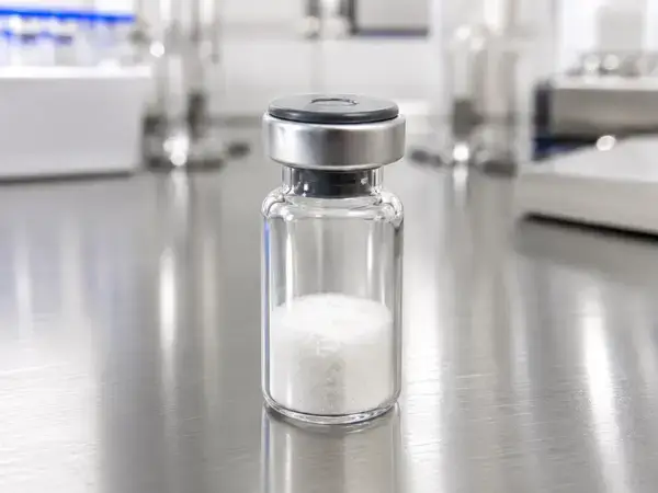
An acylated GLP-1 analog carries a lipid (fatty acid) chain attached to a lysine residue on the peptide backbone, usually through a short spacer such as a gamma-glutamic acid unit and one or more PEG-style linkers. Representative analogs described in the published literature carry chains of differing length, including a C16 palmitoyl chain, a C18 fatty diacid, and a C20 fatty diacid.
That lipid tail is the source of both the analog’s defining properties and its handling challenges. Pairing a water-loving peptide backbone with a strongly water-avoiding fatty chain produces an amphipathic molecule, and amphipathic molecules want to organize themselves. In solution they cluster their lipid tails together, crowd toward interfaces, and stick to surfaces. Understanding that single fact explains most of what goes wrong during handling.
References to specific analogs here are made only in a scientific, descriptive context. This content is not affiliated with, endorsed by, or sponsored by any pharmaceutical manufacturer, and it makes no comparison to or claim about any commercial product.
Why Aggregation Happens
Aggregation in acylated GLP-1 analogs is driven by several overlapping mechanisms, and recognizing them helps you design conditions that keep material monomeric and usable.
The first driver is concentration-dependent self assembly. Above a threshold concentration, the lipid tails associate and the molecules form micelle-like structures and higher order oligomers. Push the concentration too high during reconstitution and you encourage this behavior immediately.
The second driver is interfacial stress. Amphipathic molecules migrate to air and water boundaries and to container walls, where they partially unfold and seed further aggregation. Anything that increases interfacial area, such as vigorous shaking, vortexing, or foaming, accelerates the problem.
The third driver is backbone misfolding. Peptide chains can stack into beta-sheet-rich structures and progress toward fibrils, especially under prolonged stress or unfavorable conditions.
Environmental factors tie these mechanisms together. A pH close to the molecule’s isoelectric point lowers solubility, high ionic strength can screen stabilizing charges, elevated temperature increases hydrophobic collapse, and repeated freezing and thawing concentrates solutes while creating disruptive ice interfaces.
How to Recognize Aggregation Early
Catch aggregation before it ruins an experiment by watching for both visible and analytical signals.
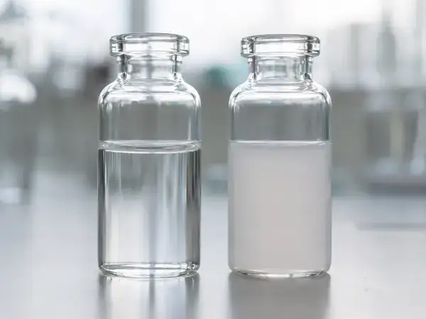
Visually, inspect every reconstituted vial against light. Cloudiness, opalescence, fine particulates, visible fibers, or a gel-like consistency all point to aggregation or precipitation. A solution that should be clear and is not should never go into a critical assay.
Analytically, several methods give you a quantitative read. Size exclusion chromatography separates monomer from oligomers and aggregates and is a workhorse for this class. Dynamic light scattering reports the size distribution of species in solution and flags early oligomerization. Reverse phase HPLC confirms purity and monomer content, and a simple turbidity reading in the near-UV range offers a fast screen. For suspected fibrils, microscopy and thioflavin-based assays are useful follow ups.
Reconstitution Best Practices
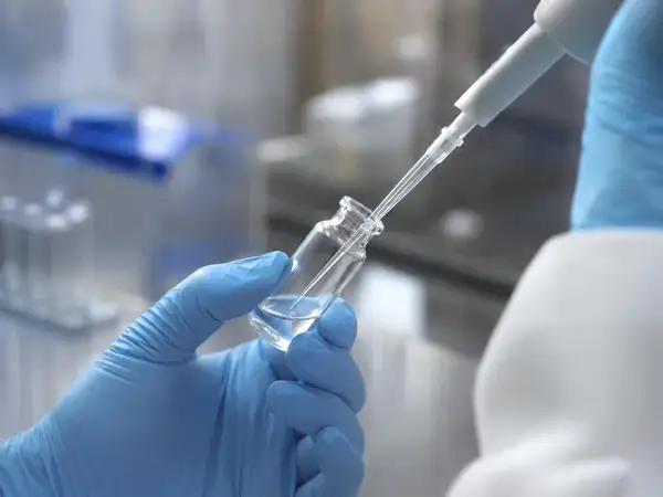
Reconstitution is where most damage happens, and a gentle, deliberate technique prevents the majority of problems. Treat the following as a standard workflow for preparing research stock solutions, and adapt the diluent and concentration to your experimental protocol.
- Equilibrate the sealed vial to room temperature before opening it. Opening a cold vial draws moisture onto the hygroscopic powder and starts degradation before you have even added diluent.
- Tap or briefly centrifuge the vial so the lyophilized material settles at the bottom.
- Select a diluent appropriate to your assay. Sterile water or a suitable buffer is common, and your downstream protocol should drive this choice.
- Add the diluent slowly, letting it run down the inner wall of the vial rather than firing it directly onto the powder. This limits local supersaturation and foaming.
- Cap the vial and mix by gentle swirling or slow inversion. Do not vortex, shake hard, or pipette aggressively. Shear forces and foam nucleate aggregation.
- Give the material a few minutes to dissolve fully, with occasional gentle swirling. Avoid heating to speed things up.
- Inspect for clarity. For analytical work, filter through a 0.22 micron membrane if your protocol calls for it, and account for possible adsorptive loss on the filter.
- For dilute working stocks, a carrier such as BSA or a buffer optimized for your assay can reduce surface losses. Confirm compatibility with your downstream method first.
Storing Reconstituted Material
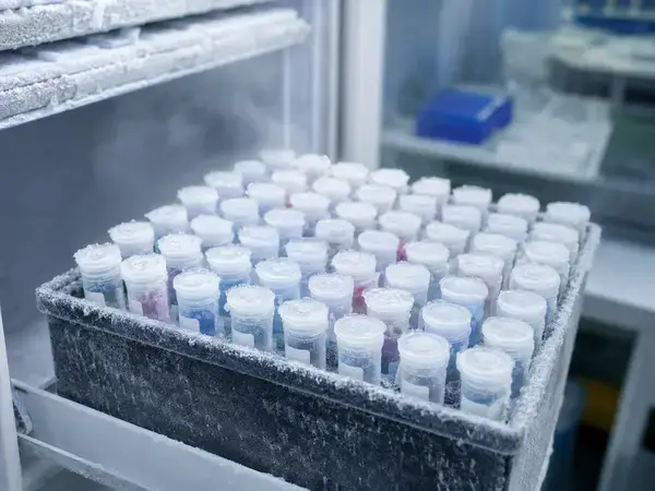
Storage decisions determine how long a stock stays useful. Keep lyophilized material cold, sealed, desiccated, and protected from light, following the storage temperature listed in your supplier’s documentation. Once reconstituted, divide the solution into single-use aliquots so you never thaw the whole batch repeatedly.
Repeated freezing and thawing is one of the most reliable ways to destroy an acylated peptide stock. Each freeze-thaw cycle concentrates solute and forms ice interfaces that drive aggregation, so minimize cycles wherever possible. Working solutions generally have limited stability, so prepare them fresh when your schedule allows and discard material that no longer passes a clarity check.
Build Good Research Records
Reliable data depends on knowing exactly what is in the vial. Verify identity and purity against the certificate of analysis that accompanies the material, paying attention to HPLC purity and mass spectrometry identity confirmation. Record lot numbers, reconstitution dates, diluents, concentrations, and storage conditions for every stock you prepare. Before any critical experiment, consider rechecking the stock by size exclusion chromatography or HPLC so you are not building results on degraded material. Consistent documentation also makes troubleshooting far easier when a result looks unexpected.
Common Pitfalls to Avoid
- Vortexing or shaking to dissolve material, which drives foaming and aggregation.
- Adding diluent directly onto the powder at high speed instead of down the vial wall.
- Opening a cold vial before it reaches room temperature.
- Storing working solutions at room temperature or subjecting them to repeated freeze and thaw cycles.
- Ignoring cloudiness or particulates and using questionable stock anyway.
- Skipping certificate of analysis review and quality checks before key experiments.
Conclusion
Acylated GLP-1 analogs reward careful handling and punish shortcuts. Treat every vial as the amphipathic, aggregation-prone molecule it is, and build your workflow around that reality. Equilibrate before opening, add diluent slowly down the wall, mix with gentle swirling instead of a vortex, and never let foam form. Store lyophilized material cold and desiccated, split reconstituted stock into single-use aliquots, and protect it from repeated freeze-thaw cycles. Confirm identity and purity against the certificate of analysis, log every reconstitution, and recheck stocks by size exclusion chromatography or HPLC before any experiment that matters. When you make these habits routine, you remove a major source of variability and protect the integrity of your data. Approach these compounds with discipline, document each step, and your research material will stay monomeric, reliable, and ready when you need it. Remember that all of this handling applies to research use only and never to human consumption.
FAQs
How can I prevent an acylated GLP-1 analog from aggregating?
Control the conditions that drive self association. Reconstitute gently by adding diluent down the vial wall and swirling rather than vortexing, keep concentrations within the range your protocol supports, and avoid foaming and hard agitation. Store the lyophilized powder cold and desiccated, aliquot reconstituted material for single use, and limit freeze-thaw cycles. Run a clarity check and an analytical method such as size exclusion chromatography before any critical work.
Why did my reconstituted solution turn cloudy?
Cloudiness usually signals aggregation or precipitation. Common causes include vortexing or shaking, adding diluent too quickly, too high a concentration, an unfavorable pH or ionic strength, temperature stress, and repeated freeze-thaw. Discard cloudy stock rather than using it, then reconstitute a fresh vial slowly and gently, confirm your diluent matches the protocol, and store the new solution in single-use aliquots.
What diluent should I use for reconstitution?
Let your experimental protocol and the supplier documentation guide the choice. Sterile water or a buffer matched to your assay is typical, and the diluent should be compatible with your downstream method. Add it slowly down the inner wall and mix gently. This guidance applies to laboratory research only and never to human or animal use.
How long does a reconstituted working solution stay usable?
Working solutions of acylated peptides generally have limited stability, so plan to use them quickly and prepare fresh material when your schedule allows. Keep aliquots cold per the supplier documentation, protect them from light, and run a clarity check before each use. If a solution shows cloudiness or particulates, discard it and reconstitute again.
Can I freeze and thaw the solution more than once?
Avoid it wherever possible. Each freeze-thaw cycle concentrates solute and creates ice interfaces that drive aggregation, which degrades the stock and adds variability to your data. Divide reconstituted material into single-use aliquots at the start so each tube is thawed only once, and discard any aliquot that fails a clarity or analytical check.