❌ Not for Human Consumption
●
✅ For Research Use Only
●
All products are intended strictly for laboratory research purposes only
●
❌ Not for Human Consumption
●
✅ For Research Use Only
●
All products are intended strictly for laboratory research purposes only
●

BPC 157 Pentadecapeptide Structure For Researchers

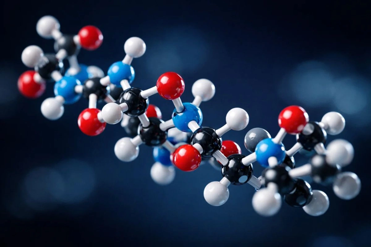
Fifteen amino acids. That’s all it takes to build one of the more structurally peculiar peptides in gastric juice research – and yet BPC 157’s compact chain raises questions that have occupied biochemists for over two decades. What makes this specific sequence so resistant to the enzymatic degradation that dismantles most peptides of comparable length within minutes? The answer sits in its molecular architecture.
This article breaks down BPC 157’s primary structure, its physicochemical properties, and the specific features that make it an unusual candidate for stability studies in peptide chemistry. If you work with synthetic peptides in a research setting, the structural details here should sharpen your understanding of why this particular compound keeps appearing in the literature.
Disclaimer: BPC 157 is sold strictly for in vitro research and laboratory use only. It is not intended for human consumption, therapeutic application, or diagnostic use. Nothing in this article constitutes medical advice or a claim of therapeutic efficacy. All researchers must comply with applicable federal, state, and institutional regulations when handling research peptides.
The Primary Sequence and Its Origins
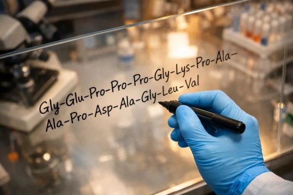
BPC 157 is a partial sequence derived from a larger protein found in human gastric juice. Its 15-residue chain follows this order:
Gly-Glu-Pro-Pro-Pro-Gly-Lys-Pro-Ala-Asp-Asp-Ala-Gly-Leu-Val
The molecular formula is C62H98N16O22, yielding a molecular weight of approximately 1,419.53 g/mol. For researchers running mass spectrometry, the expected monoisotopic mass lands near 1,418.70 Da – a useful checkpoint when verifying synthesis purity.
What immediately stands out in that sequence is the proline cluster. Three consecutive proline residues (positions 3-5) plus an additional proline at position 8 means that roughly 27% of the entire chain is proline. That’s not normal. Most bioactive peptides in the 10-20 residue range carry one or two prolines at most. This density creates structural rigidity in exactly the region where most short peptides fold loosely and expose themselves to proteolytic cleavage.
Why the Proline Content Matters for Stability Research
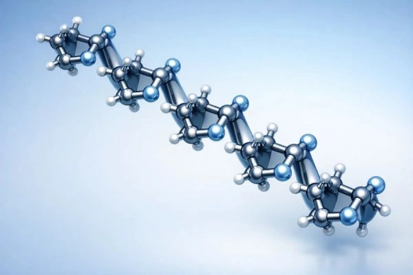
Proline is the only standard amino acid whose side chain connects back to the backbone nitrogen, forming a five-membered pyrrolidine ring. This cyclic constraint does two things that researchers studying peptide degradation care about.
First, it restricts phi-angle rotation to approximately -75 degrees, locking the backbone into conformations that resist the extended conformations most endopeptidases recognize as substrates. Second, consecutive prolines tend to form polyproline II (PPII) helices – left-handed helical structures with roughly 3.0 residues per turn and no intramolecular hydrogen bonding. PPII helices are unusually rigid for their size.
In BPC 157, the Pro-Pro-Pro segment at positions 3-5 almost certainly adopts this PPII conformation. Published circular dichroism data on comparable polyproline-rich sequences consistently show the characteristic negative band near 205 nm and positive band near 228 nm that fingerprint PPII structure. This isn’t speculative – it’s a direct consequence of stereochemistry.
The practical implication for researchers: this proline-driven rigidity likely explains why BPC 157 retains structural integrity in gastric-juice-like conditions (pH 2-3, pepsin-rich) where peptides of similar molecular weight typically degrade within 15-30 minutes. The PPII helix simply doesn’t present the flexible, extended backbone conformations that pepsin’s binding cleft requires.
Charge Distribution and Solubility Profile
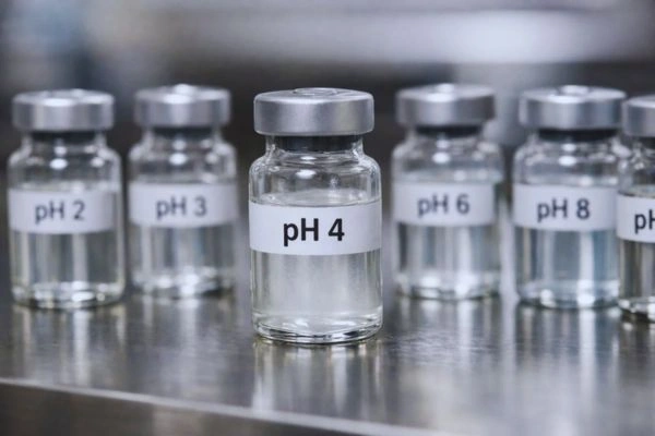
At physiological pH (7.4), BPC 157 carries a net negative charge. The two aspartate residues (positions 10-11) and one glutamate (position 2) contribute three negative charges, while lysine at position 7 provides the sole positive charge. The calculated isoelectric point sits near pH 4.2.
This charge distribution has direct consequences for researchers working with BPC 157 in solution.
In acidic buffers below pH 3, the peptide approaches net neutrality as the carboxylate groups protonate. Between pH 4 and 6, it transitions through its isoelectric point – the zone of minimum solubility where aggregation risk peaks. Above pH 7, the peptide is highly soluble due to the three deprotonated acidic residues.
For practical dissolution in laboratory settings, most protocols call for reconstitution in bacteriostatic water or sterile saline at neutral to slightly acidic pH. The peptide’s solubility in aqueous media exceeds 10 mg/mL under these conditions, making it straightforward to prepare stock concentrations for in vitro assays. Researchers should avoid reconstitution near pH 4.2, where precipitation becomes likely even at low concentrations.
Structural Comparison with Other Gastric Peptides
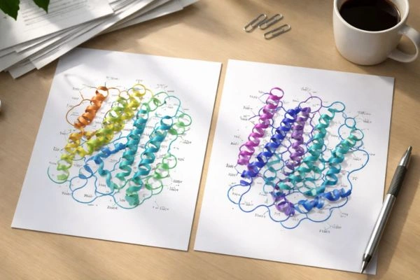
What separates BPC 157 from other well-characterized gastric peptides isn’t just the proline content – it’s the combination of proline density, small size, and the absence of disulfide bonds.
Consider gastrin-17, a 17-residue peptide also found in the gastric environment. Gastrin achieves its structural stability partly through a pyroglutamate cap at the N-terminus and C-terminal amidation. BPC 157 uses neither of these modifications. Its stability appears to be entirely sequence-driven rather than relying on post-translational modifications.
Or compare it with secretin, a 27-residue peptide that adopts an alpha-helical structure stabilized by hydrophobic packing. BPC 157’s three glycine residues (positions 1, 6, and 13) would normally destabilize helical structures – glycine is a well-documented helix-breaker due to its conformational flexibility. The peptide doesn’t appear to rely on alpha-helical organization at all.
This makes BPC 157 an interesting model compound for researchers studying how primary sequence alone – without disulfide bonds, capping groups, or helical secondary structure – can confer resistance to proteolytic degradation.
Considerations for Synthesis and Quality Control
Researchers sourcing BPC 157 for laboratory work should be aware of several synthesis-related structural details that affect experimental reproducibility.
The peptide exists in two common salt forms: BPC 157 acetate and BPC 157 sodium salt. The acetate form typically shows higher purity by HPLC (often exceeding 98%) and is the more commonly supplied research-grade form. However, the counterion affects both the actual peptide content per milligram and the buffer capacity when dissolved. A 1 mg vial of BPC 157 acetate contains less active peptide by mass than 1 mg of the free acid form. Researchers calculating molar concentrations need to account for this discrepancy.
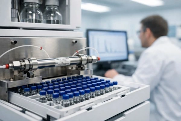
Verification of synthesis quality should include at minimum three analytical checks: HPLC purity assessment (C18 reverse-phase, acetonitrile/water gradient with 0.1% TFA), mass spectrometry confirmation of the expected molecular weight, and amino acid analysis to verify the correct residue ratios. The triple-proline segment can create challenges during solid-phase synthesis, particularly at the Glu-Pro junction (positions 2-3), where coupling efficiency sometimes drops below 95%. Incomplete coupling at this position produces a des-Pro deletion peptide with a mass approximately 97 Da lighter than the target.
Researchers receiving BPC 157 from any supplier should request certificates of analysis that include all three of these verification methods. A single HPLC chromatogram alone isn’t sufficient to confirm sequence identity.
BPC 157 Storage and Handling for Research Integrity
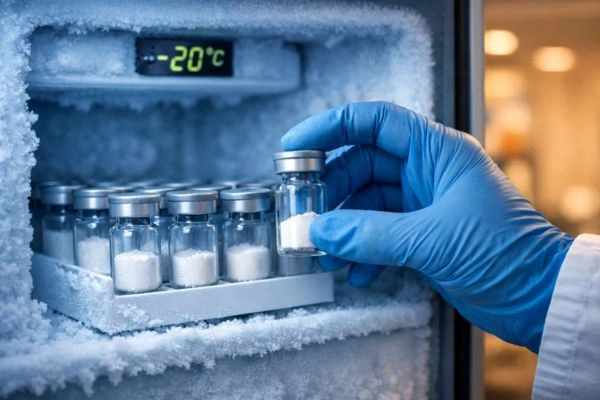
Lyophilized BPC 157 is remarkably stable when stored properly – published data suggests minimal degradation over 24 months at -20 degrees Celsius in sealed, desiccated vials. The peptide’s lack of cysteine residues means oxidative degradation pathways that plague disulfide-containing peptides don’t apply here.
Once reconstituted, the stability window narrows considerably. In aqueous solution at 4 degrees Celsius, the peptide maintains structural integrity for approximately 7-14 days, depending on pH, concentration, and the presence of preservatives like benzyl alcohol. At room temperature, hydrolysis of the Asp-Asp bond (positions 10-11) becomes the primary degradation pathway, with detectable cleavage products appearing within 48-72 hours.
For researchers running longitudinal assays, aliquoting reconstituted stock into single-use volumes and storing at -20 degrees Celsius extends usable shelf life to approximately 30 days. Repeated freeze-thaw cycles should be limited to three or fewer, as each cycle risks both peptide aggregation and adsorption losses to container walls. Low-bind polypropylene tubes reduce adsorption compared to standard polypropylene or glass.

Where This Leaves Researchers
BPC 157’s structural profile – the unusual proline density, the PPII helical propensity, the charge-driven solubility behavior, and the sequence-dependent proteolytic resistance – makes it a genuinely distinctive model peptide for several lines of investigation. Its relevance extends to fundamental questions in peptide chemistry: how does primary sequence govern stability in harsh enzymatic environments, and can those principles inform the design of other protease-resistant research compounds?
The 15-residue chain packs more structural complexity than its modest molecular weight suggests. For researchers working with it, understanding these molecular properties isn’t optional background reading – it’s the foundation for designing experiments that produce reproducible, interpretable data.
Conclusion
Every experimental design choice involving BPC 157 traces back to that 15-residue sequence. The four prolines dictating PPII helix formation, the Asp-Asp bond at positions 10-11 serving as the primary hydrolysis vulnerability in solution, the isoelectric point at pH 4.2 marking the aggregation danger zone – these aren’t academic details. They’re the variables that determine whether your assay data means anything.
Researchers entering this space should prioritize three things immediately: source only from suppliers providing triple-verified COAs (HPLC, mass spec, and amino acid analysis), reconstitute exclusively above pH 5 to avoid precipitation artifacts, and aliquot into single-use low-bind tubes on day one. Skip any of those steps and you’re introducing confounds that no statistical correction can fix.
The peptide’s real value to the field isn’t what it does – it’s what its architecture teaches us about sequence-driven protease resistance in miniature chains.
Disclaimer: BPC 157 is sold strictly for in vitro research and laboratory use only. It is not intended for human consumption, therapeutic application, or diagnostic use. Nothing in this article constitutes medical advice or a claim of therapeutic efficacy. All researchers must comply with applicable federal, state, and institutional regulations when handling research peptides.
Frequently Asked Questions
What is BPC 157’s amino acid sequence and molecular weight?
The sequence runs Gly-Glu-Pro-Pro-Pro-Gly-Lys-Pro-Ala-Asp-Asp-Ala-Gly-Leu-Val, with a molecular formula of C62H98N16O22 and a molecular weight of approximately 1,419.53 g/mol. When running mass spectrometry for purity verification, target a monoisotopic mass near 1,418.70 Da. Confirm this value against your supplier’s certificate of analysis before proceeding with any assay work.
Why is BPC 157 resistant to enzymatic degradation in gastric conditions?
Four proline residues – three consecutive at positions 3-5, plus one at position 8 – lock the backbone into a polyproline II helix that pepsin’s binding cleft can’t accommodate. Unlike gastrin-17 or secretin, BPC 157 achieves this stability without disulfide bonds, N-terminal capping, or C-terminal amidation. The resistance is purely sequence-driven, which is what makes it a useful model compound for protease-resistance research.
What’s the difference between BPC 157 acetate and BPC 157 sodium salt?
The acetate form is the more common research-grade option, typically exceeding 98% purity by HPLC. The critical operational difference: acetate salt adds counterion mass, so 1 mg of BPC 157 acetate contains less active peptide than 1 mg of the free acid. Adjust your molar concentration calculations accordingly, or you’ll systematically underdose every in vitro assay. Request the peptide content percentage from your supplier’s COA to calculate accurately.
How should researchers store and handle reconstituted BPC 157?
Lyophilized powder holds stable for 24+ months at -20°C in sealed, desiccated vials. Once dissolved, the window shrinks fast – 7-14 days at 4°C before the Asp-Asp bond at positions 10-11 begins hydrolyzing. Aliquot your reconstituted stock into single-use low-bind polypropylene tubes on the day of preparation and store at -20°C for up to 30 days. Cap freeze-thaw cycles at three maximum. Standard polypropylene and glass both cause measurable adsorption losses.
What quality control checks should researchers require from BPC 157 suppliers?
Demand three things on every certificate of analysis: C18 reverse-phase HPLC purity data (acetonitrile/water gradient, 0.1% TFA), mass spectrometry confirming the target molecular weight, and amino acid analysis verifying correct residue ratios. A single HPLC chromatogram alone can’t distinguish BPC 157 from its most common synthesis byproduct – a des-Pro deletion peptide roughly 97 Da lighter, caused by incomplete coupling at the Glu-Pro junction. If a supplier only provides HPLC, find a different supplier.