
NAD+ In Cellular Energy Research: A Review Of Published In Vitro And In Vivo Studies

Research Use Notice: NAD+ products referenced in this article are intended exclusively for in vitro and in vivo laboratory research. They are not approved for human consumption, diagnostic use, or therapeutic application.
Nicotinamide adenine dinucleotide (NAD+) stands as one of the most extensively studied coenzymes in modern biochemistry. Since its initial discovery in the early 20th century, NAD+ has remained at the center of cellular energy metabolism research, attracting the sustained attention of molecular biologists, biochemists, and cellular physiologists worldwide.
The growing body of published in vitro and in vivo studies exploring NAD+ has expanded our understanding of its fundamental roles in oxidative phosphorylation, glycolysis, the tricarboxylic acid (TCA) cycle, and a host of enzymatic reactions critical to cellular function. For professional researchers seeking high-purity NAD+ for laboratory investigation, understanding the existing literature is essential to designing rigorous experiments and contributing meaningful data to the field.
This article provides a structured review of the published scientific literature on NAD+ as it relates to cellular energy research, summarizing key findings from both in vitro (cell-based) and in vivo (animal model) studies. All information is presented for educational purposes and is intended to serve the professional research community.
Disclaimer: The NAD+ referenced throughout this article is sold strictly for research purposes only and is not intended for human consumption. This content is provided for educational and informational purposes directed at professional researchers, scientists, and academic institutions. Nothing in this article constitutes medical advice, a therapeutic claim, or an endorsement of any unapproved use. All referenced studies are cited from peer-reviewed literature to support the academic understanding of NAD+ biochemistry.

Section 1: The Biochemical Foundation of NAD+ in Cellular Energy
1.1 Structure and Redox Function
NAD+ is a dinucleotide composed of two nucleotides joined through their phosphate groups. One nucleotide contains an adenine nucleobase and the other contains nicotinamide. In its oxidized form (NAD+), the molecule serves as a primary electron acceptor in catabolic reactions. Upon accepting a hydride ion (two electrons and one proton), it is reduced to NADH.
This redox cycling between NAD+ and NADH is foundational to cellular bioenergetics. Published biochemistry literature consistently identifies this interconversion as a rate-limiting factor in several metabolic pathways (Cantó et al., 2015; Verdin, 2015).
1.2 NAD+ in Core Metabolic Pathways
Research has established that NAD+ participates directly in three central metabolic pathways:
Glycolysis: During glycolysis, NAD+ functions as an electron acceptor in the reaction catalyzed by glyceraldehyde-3-phosphate dehydrogenase (GAPDH). Published kinetic studies have demonstrated that the availability of cytosolic NAD+ can directly influence glycolytic flux in isolated cell preparations (Williamson et al., 1967).
The TCA Cycle: Within the mitochondrial matrix, NAD+ serves as the electron acceptor for three dehydrogenase reactions in the TCA cycle: isocitrate dehydrogenase, alpha-ketoglutarate dehydrogenase, and malate dehydrogenase. In vitro reconstitution experiments have confirmed the stoichiometric requirement of NAD+ for cycle progression (Hansford, 1983).
Oxidative Phosphorylation: NADH generated by glycolysis and the TCA cycle donates electrons to Complex I (NADH: ubiquinone oxidoreductase) of the electron transport chain (ETC). The regeneration of NAD+ at this step is essential for maintaining metabolic throughput, a finding consistently replicated across multiple in vitro mitochondrial isolation studies (Chance & Williams, 1955).
1.3 NAD+ as a Substrate for Non-Redox Enzymes
Beyond its redox function, NAD+ serves as a consumed substrate for several enzyme families of significant research interest:
Sirtuins (SIRTs): This family of NAD+-dependent deacylases cleaves NAD+ during their catalytic cycle. In vitro enzymatic assays have demonstrated that sirtuin activity is directly proportional to NAD+ availability within physiologically relevant concentration ranges (Imai et al., 2000).
Poly(ADP-ribose) Polymerases (PARPs): PARPs consume NAD+ during the process of poly(ADP-ribosyl)ation, a post-translational modification involved in DNA damage response. Published studies using recombinant PARP proteins have shown that sustained PARP activation can significantly deplete intracellular NAD+ pools in cultured cell models (Bai & Cantó, 2012).
CD38/CD157 Ectoenzymes: These glycohydrolases catalyze the degradation of NAD+ to produce cyclic ADP-ribose. Research using CD38 knockout animal models has provided significant data regarding the relationship between CD38 activity and tissue NAD+ levels (Camacho-Pereira et al., 2016).
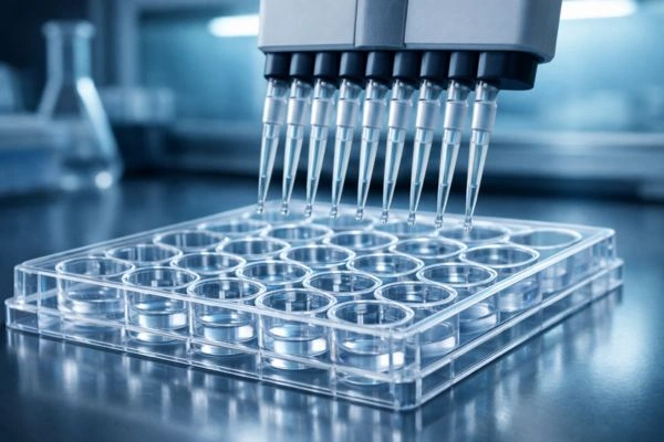
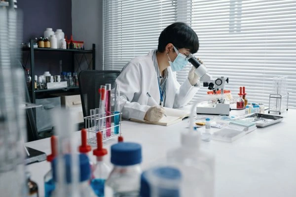
Section 2: Review of Published In Vitro Studies
In vitro studies have been instrumental in isolating specific mechanisms by which NAD+ influences cellular energy production. The following subsections review key published findings from cell-free systems and cultured cell experiments.
2.1 Mitochondrial Respiration Assays
A substantial body of literature has utilized isolated mitochondria preparations to study the effect of NAD+ on respiratory chain function. Classic experiments by Chance and Williams (1955) established the relationship between NAD+/NADH ratios and mitochondrial respiratory states. More recent studies using high-resolution respirometry (e.g., Oroboros O2k systems) have refined these measurements, demonstrating that supplementation of NAD+ to permeabilized cell preparations can restore respiratory capacity in models of metabolic dysfunction (Pirinen et al., 2020).
2.2 NAD+ Biosynthesis Pathway Studies in Cell Culture
Researchers have used cultured mammalian cell lines to map the relative contributions of different NAD+ biosynthesis pathways. Published studies in HEK293 cells and primary hepatocytes have quantified flux through the de novo synthesis pathway (from tryptophan), the Preiss-Handler pathway (from nicotinic acid), and the salvage pathway (from nicotinamide). Tracer studies using isotopically labeled precursors have revealed that the salvage pathway, catalyzed by nicotinamide phosphoribosyltransferase (NAMPT), is the dominant route for NAD+ regeneration in most mammalian cell types under standard culture conditions (Revollo et al., 2004; Liu et al., 2018).
2.3 NAD+ Depletion Models
To study the consequences of reduced NAD+ availability, researchers have employed pharmacological inhibitors such as FK866 (a NAMPT inhibitor) in cell culture. Published data from these experiments demonstrate that progressive NAD+ depletion leads to measurable decreases in cellular ATP content, impairment of mitochondrial membrane potential, and ultimately, cell death through necrotic and apoptotic mechanisms (Hasmann & Schemainda, 2003). These depletion models remain valuable tools for investigating the threshold concentrations of NAD+ required to sustain various aspects of cellular energy metabolism.
2.4 Sirtuin Activity Assays
Fluorometric and luminescence-based sirtuin activity assays performed in vitro have consistently demonstrated a dose-dependent relationship between NAD+ concentration and deacetylase activity for SIRT1, SIRT3, and other sirtuin family members. Studies using recombinant enzymes and acetylated peptide substrates have defined the Km values of various sirtuins for NAD+, providing essential kinetic parameters for the field (Feldman et al., 2012).
Section 3: Review of Published In Vivo Studies
While in vitro studies allow for precise mechanistic control, in vivo studies provide critical context about how NAD+ metabolism operates within complex biological systems. The following subsections summarize key findings from published animal model research.
3.1 Age-Associated Decline of NAD+ in Animal Models
One of the most consistently replicated findings in the NAD+ literature is the observation that tissue NAD+ levels decline with age in multiple animal model species. Studies in aged C57BL/6 mice have reported significant reductions in NAD+ concentrations across multiple tissues, including liver, skeletal muscle, brain, and adipose tissue, when compared to young controls (Yoshino et al., 2011; Gomes et al., 2013). These findings have established a widely referenced framework for studying the relationship between NAD+ availability and age-associated metabolic changes in preclinical models.
3.2 NAD+ Precursor Administration Studies
A large number of in vivo studies have examined the effects of administering NAD+ precursors, particularly nicotinamide riboside (NR) and nicotinamide mononucleotide (NMN), to animal models. Key published findings include:
Metabolic Parameters: Yoshino et al. (2011) reported that administration of NMN to aged mice resulted in measurable increases in tissue NAD+ levels and alterations in several metabolic biomarkers. Cantó et al. (2012) demonstrated that NR supplementation in high-fat-diet-fed mice was associated with changes in mitochondrial content in skeletal muscle and brown adipose tissue, as assessed by mitochondrial DNA copy number and citrate synthase activity.
Mitochondrial Function Markers: Multiple studies have assessed mitochondrial function endpoints following NAD+ precursor administration in animal models. Published data include measurements of oxygen consumption rates in isolated tissue mitochondria, expression levels of ETC complex subunits, and mitochondrial membrane potential indicators (Mills et al., 2016; Zhang et al., 2016).
Exercise Physiology Models: Frederick et al. (2016) published data from a murine exercise model demonstrating that NMN administration was associated with measurable changes in exercise endurance parameters and skeletal muscle NAD+ levels in treated versus control groups.
3.3 Genetic Models of NAD+ Metabolism
Transgenic and knockout animal models have provided critical mechanistic data. Notable published studies include:
NAMPT Tissue-Specific Knockouts: Researchers have generated conditional knockout models to study the effects of ablating NAD+ salvage biosynthesis in specific tissues. Published data from skeletal muscle-specific NAMPT knockout mice demonstrated significant impairment of muscle mitochondrial function and exercise capacity (Frederick et al., 2016).
CD38 Knockout Models: As referenced above, CD38 knockout mice exhibit elevated tissue NAD+ levels compared to wild-type controls. Published studies in these models have been widely cited in the literature as evidence supporting the role of CD38 as a major NAD+ consuming enzyme in vivo (Camacho-Pereira et al., 2016).
PARP1 Knockout Models: Studies in PARP1-deficient mice have reported elevated basal NAD+ levels in multiple tissues and have been used to investigate the relationship between PARP activity, NAD+ availability, and mitochondrial function (Bai et al., 2011).
3.4 Tissue-Specific NAD+ Measurements
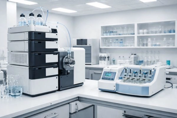
Advances in analytical chemistry, particularly liquid chromatography-mass spectrometry (LC-MS) methods, have enabled researchers to perform precise quantification of NAD+ and its metabolites across different tissues in animal models. Published methodological studies have validated protocols for extracting and measuring NAD+, NADH, NMN, NR, and related metabolites from flash-frozen tissue samples (Trammell & Brenner, 2013). These analytical methods are essential tools for any research laboratory investigating NAD+ metabolism, and the availability of high-purity NAD+ reference standards is critical for accurate quantitation.
Section 4: Current Research Frontiers
The following areas represent active frontiers in NAD+ research as reflected in the recently published literature. These are noted here to provide professional researchers with context for ongoing and future investigations.
4.1 NAD+ Compartmentalization
Recent studies have highlighted the importance of subcellular NAD+ compartmentalization. Research using genetically encoded NAD+ biosensors has revealed that mitochondrial, nuclear, and cytosolic NAD+ pools are regulated semi-independently and may respond differently to metabolic perturbations (Cambronne et al., 2016). Understanding compartment-specific NAD+ dynamics remains an active area of investigation.
4.2 NAD+ and Circadian Rhythm Research
Published studies have identified a reciprocal relationship between NAD+ metabolism and circadian clock machinery. NAMPT expression, and consequently NAD+ biosynthesis, follows a circadian oscillation pattern in murine liver tissue. Sirtuin-mediated feedback on clock gene expression creates a regulatory loop linking NAD+ availability to circadian biology (Nakahata et al., 2009; Ramsey et al., 2009).
4.3 NAD+ Metabolomics and Flux Analysis
The application of stable isotope tracing and metabolic flux analysis to NAD+ metabolism represents a growing area of research. Published studies using deuterium- and carbon-13-labeled precursors have begun to quantify the rates of NAD+ synthesis, consumption, and recycling in both in vitro and in vivo systems (Liu et al., 2018). These approaches require high-purity reagents, including NAD+ standards, for accurate experimental design and data interpretation.
4.4 Intercellular NAD+ Metabolism
Emerging research has begun to investigate NAD+ metabolism at the intercellular and systemic level, exploring how NAD+ intermediates may be exchanged between cells and tissues. Published studies have identified extracellular NAMPT (eNAMPT) and circulating NMN as potential mediators of inter-tissue metabolic communication in animal models (Yoshida et al., 2019).
Section 5: Considerations for Laboratory Researchers
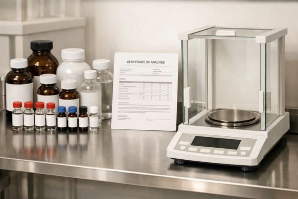
5.1 Purity and Quality of NAD+ for Research Use
The reliability and reproducibility of NAD+ research depend significantly on the purity and quality of reagents used. Researchers should ensure that NAD+ used in experimental protocols meets established analytical standards, including verification by HPLC, mass spectrometry, or enzymatic activity assays. Certificates of analysis (COAs) should accompany all research-grade NAD+ products.
5.2 Stability and Storage
Published stability studies indicate that NAD+ is sensitive to degradation by heat, light, and extreme pH. Researchers should store NAD+ under conditions recommended by the supplier, typically at or below -20°C in a desiccated environment, to maintain integrity for experimental use.
5.3 Experimental Controls
As with any bioactive reagent, proper experimental controls are essential. Published best practices recommend including vehicle-only controls, dose-response analyses, and orthogonal validation methods when investigating NAD+-dependent processes in both in vitro and in vivo systems.
Conclusion
The published scientific literature on NAD+ in cellular energy research spans decades of rigorous investigation and continues to expand rapidly. From foundational biochemical studies establishing its role in glycolysis, the TCA cycle, and oxidative phosphorylation, to contemporary in vivo studies utilizing advanced genetic models and metabolomics techniques, NAD+ remains a molecule of central importance to our understanding of cellular bioenergetics.
For professional researchers and academic investigators, access to high-purity, research-grade NAD+ is an essential requirement for contributing to this growing body of knowledge. The studies reviewed in this article represent only a fraction of the available literature, and researchers are encouraged to consult primary sources and current review articles when designing their experimental protocols.
Disclaimer: The NAD+ referenced throughout this article is sold strictly for research purposes only and is not intended for human consumption. This content is provided for educational and informational purposes directed at professional researchers, scientists, and academic institutions. Nothing in this article constitutes medical advice, a therapeutic claim, or an endorsement of any unapproved use. All referenced studies are cited from peer-reviewed literature to support the academic understanding of NAD+ biochemistry.
Frequently Asked Questions
Why is NAD+ considered essential for cellular energy metabolism research?
NAD+ directly participates in glycolysis, the TCA cycle, and oxidative phosphorylation as a primary electron acceptor, making it a rate-limiting factor in ATP production. It also serves as a consumed substrate for sirtuins, PARPs, and CD38, enzyme families central to metabolic regulation and DNA repair. Researchers studying bioenergetics at any level should account for NAD+ availability as a fundamental experimental variable.
What are the most widely used in vitro models for studying NAD+ function?
Common approaches include isolated mitochondria respiration assays, cultured mammalian cell lines (such as HEK293 and primary hepatocytes) for biosynthesis pathway mapping, pharmacological depletion models using NAMPT inhibitors like FK866, and fluorometric sirtuin activity assays with recombinant enzymes. Select the model that best aligns with your specific research question, whether it involves respiratory chain function, pathway flux, or enzyme kinetics.
How do researchers measure NAD+ levels accurately in tissue samples?
Liquid chromatography–mass spectrometry (LC-MS) is the current gold standard for quantifying NAD+ and its metabolites in flash-frozen tissue. Validated extraction protocols allow simultaneous measurement of NAD+, NADH, NMN, NR, and related intermediates. Ensure your laboratory uses high-purity NAD+ reference standards and follows published sample-handling procedures to maintain measurement accuracy.
What purity and storage standards should research-grade NAD+ meet?
Research-grade NAD+ should be verified by HPLC, mass spectrometry, or enzymatic activity assays, and each lot should be accompanied by a certificate of analysis (COA). Store NAD+ at or below −20°C in a desiccated environment, as the molecule is sensitive to degradation by heat, light, and extreme pH. Always confirm reagent integrity before use in time-sensitive or resource-intensive experiments.
What are the current frontiers in NAD+ research that laboratories should consider exploring?
Active research areas include subcellular NAD+ compartmentalization using genetically encoded biosensors, the reciprocal link between NAD+ metabolism and circadian clock regulation, stable isotope–based metabolic flux analysis, and intercellular NAD+ communication mediated by extracellular NAMPT and circulating NMN. These frontiers offer high-impact opportunities for laboratories equipped with advanced analytical and genetic tools.