❌ Not for Human Consumption
●
✅ For Research Use Only
●
All products are intended strictly for laboratory research purposes only
●
❌ Not for Human Consumption
●
✅ For Research Use Only
●
All products are intended strictly for laboratory research purposes only
●

Recombinant Expression And Purification Of IGF-1 Analogs For Laboratory Use


Written By: Gary Hite, Research Content Writer
Reviewed By: Natalie Kunsman, M.D., Board-Certified Physician
Last Reviewed: September 21, 2026
Insulin-like growth factor 1 (IGF-1) and its engineered analogs rank among the most widely studied polypeptides in molecular biology. For laboratories that depend on consistent, well-characterized material, recombinant expression offers a dependable route to producing these proteins at the bench. This guide walks through the practical workflow, from construct design to final polishing, so research teams can plan a production strategy that matches their experimental goals.
Disclaimer: All products and information referenced are intended strictly for research purposes only and are not for human consumption. These materials are not drugs, foods, supplements, or medical devices, and they are not intended to diagnose, treat, cure, or prevent any condition. They are sold and used exclusively by qualified professionals and academic researchers for laboratory and educational purposes, in full compliance with applicable laws and institutional guidelines.
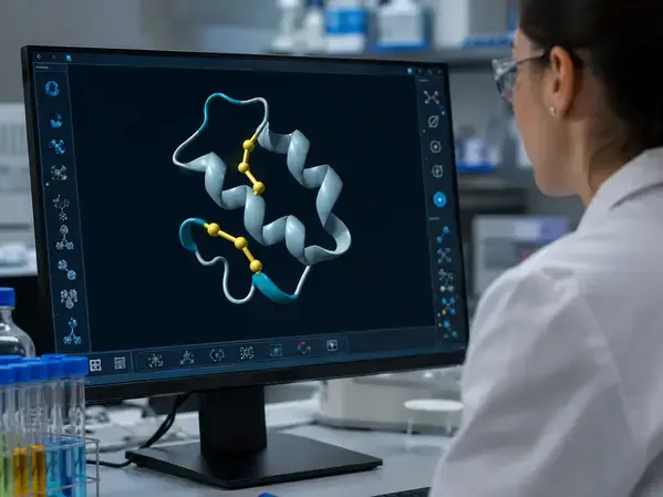
Understanding IGF-1 and Its Analogs in a Research Context
IGF-1 is a single chain polypeptide of roughly 70 amino acids, with a molecular weight near 7.6 kDa. Its compact fold is stabilized by three intramolecular disulfide bonds, and that structural detail drives most of the technical decisions in any production workflow. Correct disulfide pairing is the central challenge, because misfolded material behaves differently in downstream assays and complicates purity assessment.
Research teams study recombinant IGF-1 to explore questions in protein structure, receptor binding, and signaling biology under defined laboratory conditions. Several engineered analogs appear frequently in the published literature as reference reagents. Long R3 IGF-1 (LR3-IGF-1) carries an amino acid substitution along with a short N-terminal extension that shifts its interaction with IGF binding proteins in vitro. Des(1-3)IGF-1 lacks the first three N-terminal residues, which similarly alters its binding behavior in controlled assays. These variants give investigators a way to probe specific aspects of IGF-1 biochemistry, and they are supplied and handled as laboratory reference materials only.
Why Recombinant Expression Suits Laboratory Work
Chemical synthesis of a 70 residue polypeptide that also requires three correctly paired disulfide bonds is demanding and rarely practical at scale. Recombinant expression sidesteps those limits by letting cells assemble the protein backbone with high sequence fidelity. The approach delivers several advantages that matter for reproducible research:
- Precise control over the amino acid sequence, including defined analog mutations.
- Scalable batches that support repeat experiments with consistent lot characteristics.
- The option to add purification and solubility tags directly to the construct.
- A workflow that adapts to bacterial, yeast, or mammalian platforms as the project requires.
With those benefits in mind, the first major decision is choosing the right expression host.
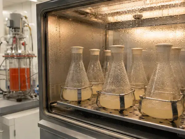
Selecting an Expression System
The host you select shapes yield, folding quality, and the complexity of every step that follows. Match the platform to the demands of your analog and your downstream assays.
Bacterial Expression
Escherichia coli remains the workhorse for IGF-1 production. It grows quickly, scales readily, and produces high protein levels. The tradeoff is that the bacterial cytoplasm is a reducing environment, so disulfide rich proteins like IGF-1 commonly accumulate as insoluble inclusion bodies. That outcome is manageable and, in many cases, even useful, because inclusion bodies concentrate the target protein and protect it from proteolysis. The extra effort simply shifts later, into the refolding stage.
Yeast Expression
Pichia pastoris and related yeast hosts offer a secretory pathway with an oxidizing environment that can support disulfide formation. Secreted product simplifies the early stages of purification because much of the host protein stays inside the cell. Yeast systems suit teams that want folded material directly from the culture supernatant and are willing to invest in strain development.
Mammalian and Insect Cell Expression
Mammalian platforms such as CHO and HEK293 lines, along with baculovirus driven insect cell systems, deliver proteins through native folding and processing machinery. These hosts typically produce lower yields and require more involved culture handling, yet they are valuable when an analog must fold under conditions that closely mirror a eukaryotic setting for in vitro study.
Designing the Expression Construct
A thoughtful construct removes problems before they appear. Start by optimizing the coding sequence for the chosen host so that codon usage supports efficient translation. Pair the gene with a strong, inducible promoter. The T7 system is a common choice for E. coli work because it gives tight, high level control over expression.
Tags do a great deal of the heavy lifting. A polyhistidine tag (His6) enables straightforward affinity capture, while solubility partners such as small ubiquitin-like modifier (SUMO), thioredoxin, maltose binding protein, or glutathione S-transferase can improve folding and reduce inclusion body formation. SUMO fusions are especially popular for compact, disulfide rich targets because they aid solubility and leave a native N-terminus after cleavage.
Plan tag removal from the start. Insert a defined protease site, such as a TEV, SUMO protease, thrombin, or enterokinase recognition sequence, between the tag and the mature protein. The right placement lets you release the analog cleanly once it has served its purpose. For secretion based systems, include an appropriate signal sequence so the host directs the product to the correct compartment.
Managing Inclusion Bodies and Refolding
When E. coli deposits your analog into inclusion bodies, treat that as a defined stage rather than a setback. Begin by lysing the cells and isolating the dense inclusion body fraction through centrifugation, then wash it to strip away loosely associated contaminants.
Solubilize the washed pellet with a strong chaotrope, typically urea or guanidine hydrochloride, often together with a reducing agent to fully unfold the chains. The refolding step then coaxes the polypeptide back into its native, disulfide bonded form. Common strategies include slow dilution into a refolding buffer or stepwise dialysis to gradually lower the denaturant concentration. A redox couple such as reduced and oxidized glutathione encourages correct disulfide shuffling, and additives like L-arginine help suppress aggregation during the transition. Monitor refolding yield with analytical methods so you can tune buffer composition, pH, temperature, and protein concentration for the best recovery of correctly folded material.
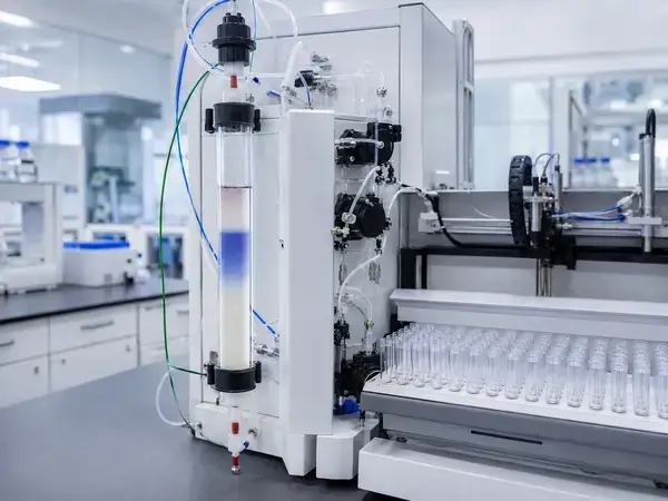
Building the Purification Workflow
A robust purification plan moves through three logical phases: capture, intermediate purification, and polishing. Each phase tightens purity and removes a different class of contaminant.
For capture, immobilized metal affinity chromatography (IMAC) on a nickel resin pairs naturally with a His-tagged construct and pulls the target out of a complex mixture in a single bind and elute step. After capture, remove the tag with your chosen protease, then separate the freed analog from the cleaved tag and the protease itself.
Intermediate purification often relies on ion exchange chromatography. Because IGF-1 carries a basic isoelectric point, cation exchange performed below that pH binds the protein efficiently and resolves it from charge variant impurities. Finish with a polishing step that delivers the high purity research applications demand. Size exclusion chromatography separates the monomer from aggregates and residual fragments, and reversed phase high-performance liquid chromatography can push purity even higher while confirming homogeneity.
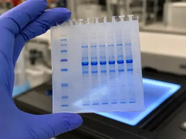
Characterization and Quality Control
Purity claims mean little without data to support them, so characterize every batch with a complementary panel of methods. The goal is a clear, documented picture of identity, purity, and structural integrity.
- SDS-PAGE to confirm the expected molecular weight and flag visible impurities.
- Reversed phase HPLC to quantify purity and detect closely related species.
- Mass spectrometry for intact mass confirmation and peptide mapping to verify sequence.
- Disulfide bond analysis to confirm correct folding, the defining quality marker for IGF-1.
- Protein concentration by absorbance at 280 nm or a colorimetric assay.
- Endotoxin testing to document low-endotoxin status for sensitive in vitro work.
Where the experimental design calls for it, in vitro binding or activity assays provide an additional functional readout under controlled laboratory conditions. Record every result in a certificate of analysis so collaborators can trace material back to a defined lot.
Handling, Storage, and Documentation
Careful handling protects the work you have already invested. Many laboratories store IGF-1 analogs as a lyophilized powder for long-term stability, then reconstitute in a suitable buffer just before use. Prepare single use aliquots to avoid repeated freeze and thaw cycles, which can degrade a disulfide rich protein over time. Follow established temperature guidelines for both the dry and the reconstituted forms, and label each aliquot with lot information so traceability stays intact across the project.
Maintain thorough records throughout. A documented chain from expression batch to final aliquot supports reproducibility, simplifies troubleshooting, and keeps your laboratory aligned with good scientific practice.
Compliance and Responsible Research Use
Producing and handling research proteins comes with clear professional responsibilities. Confirm that your laboratory follows all applicable institutional, biosafety, and regulatory guidelines for recombinant work, and that personnel are trained in the relevant procedures. Dispose of biological and chemical waste according to your institution’s protocols.
Material described in this article is supplied and intended for laboratory research and educational use by qualified professionals and academic institutions. IGF-1 and its analogs covered here are for research purposes only and are not for human consumption. They are not drugs, foods, dietary supplements, cosmetics, or medical devices, and they are not intended to diagnose, treat, cure, or prevent any condition. Nothing in this content should be read as a medical claim or as encouragement to use these materials in any manner outside a controlled research setting.
Conclusion
Recombinant expression puts reliable, well-characterized IGF-1 analogs within reach of any laboratory ready to plan the workflow with care. Start by matching the expression host to your folding needs, then design a construct that pairs efficient translation with the right purification and solubility tags. Treat inclusion bodies as a defined stage, refold with a tuned redox system, and move through capture, intermediate, and polishing chromatography to reach the purity rigorous research demands. Confirm every batch with SDS-PAGE, reversed phase HPLC, mass spectrometry, and disulfide analysis, then record each result in a clear certificate of analysis. Store aliquots properly, avoid repeated freeze and thaw cycles, and keep traceability intact from culture to vial. Follow this disciplined process and your team gains consistent material that supports reproducible, rigorous science. Always remember that these IGF-1 analogs are intended strictly for research purposes only and are not for human consumption.
FAQs
Why does recombinant IGF-1 often accumulate in inclusion bodies, and how should I handle it?
The reducing environment of the E. coli cytoplasm blocks the three disulfide bonds IGF-1 needs, so the protein frequently deposits as insoluble inclusion bodies. Treat this as a planned stage rather than a failure. Isolate and wash the inclusion body fraction, solubilize it with a chaotrope such as urea or guanidine hydrochloride, then refold by slow dilution or stepwise dialysis using a glutathione redox couple. Add L-arginine to suppress aggregation, and monitor recovery so you can fine tune pH, temperature, and protein concentration for the best yield of correctly folded material.
Which expression system is best for producing IGF-1 analogs?
Choose the host that matches your folding and yield needs. E. coli delivers high yields quickly and suits teams comfortable with a refolding step. Pichia pastoris offers a secretory, oxidizing environment that supports disulfide formation and simplifies early purification. Mammalian lines such as CHO or HEK293 provide native folding for the most demanding constructs, with lower yields and more involved culture handling. Map these tradeoffs against your project goals before you commit to a platform.
What tags improve solubility and simplify purification?
Pair a purification tag with a solubility partner to get the benefit of both. A polyhistidine (His6) tag enables clean affinity capture on nickel resin, while fusion partners such as SUMO, thioredoxin, maltose binding protein, or glutathione S-transferase improve folding and reduce inclusion body formation. SUMO fusions are a strong choice for compact, disulfide rich targets because they aid solubility and leave a native N-terminus after cleavage. Always design in a defined protease site so you can remove the tag cleanly once it has done its job.
How do I confirm the purity and correct folding of a finished batch?
Run a complementary panel rather than relying on any single test. Use SDS-PAGE to check molecular weight, reversed phase HPLC to quantify purity, and mass spectrometry for intact mass and peptide mapping. Verify disulfide bonding directly, since correct folding is the defining quality marker for IGF-1. Add endotoxin testing for sensitive in vitro work, then capture every result in a certificate of analysis so collaborators can trace material back to a defined lot.
How should recombinant IGF-1 analogs be stored and handled in the laboratory?
Protect the material you worked hard to produce. Store the protein as a lyophilized powder for long term stability, then reconstitute in a suitable buffer just before use. Prepare single use aliquots to avoid repeated freeze and thaw cycles that can degrade a disulfide rich protein, and label each aliquot with lot details so traceability stays intact. Handle and store these analogs strictly as laboratory reagents, because they are intended for research purposes only and are not for human consumption.