❌ Not for Human Consumption
●
✅ For Research Use Only
●
All products are intended strictly for laboratory research purposes only
●
❌ Not for Human Consumption
●
✅ For Research Use Only
●
All products are intended strictly for laboratory research purposes only
●

HCG Immunoassay And Detection Methods For Research Use

Written By: Gary Hite, Research Content Writer
Reviewed By: Natalie Kunsman, M.D., Board-Certified Physician
Last Reviewed: September 11, 2026
Human chorionic gonadotropin (HCG) is a heterodimeric glycoprotein and one of the most widely used model analytes in immunoassay development, which makes reliable detection a core requirement for laboratories working with HCG research samples. Whether the goal is quantification, isoform discrimination, or method validation, detection accuracy depends on selecting the right immunoassay format and understanding how antibody design shapes the result. This article walks through the principal immunoassay and detection methods for HCG, written for analytical chemists, immunologists, and academic researchers.
Disclaimer: HCG research samples are intended strictly for laboratory research and educational use only. They are not for human consumption and not for any diagnostic or therapeutic application. The information below describes analytical detection methods for qualified researchers and academic professionals.
Why Immunoassays Anchor HCG Detection
HCG is a large glycoprotein composed of a common alpha subunit and a hormone-specific beta subunit, and it carries multiple distinct antibody-binding regions, or epitopes. This structure makes it well suited to antibody-based detection, where the specificity of the antibody-antigen interaction supplies both sensitivity and selectivity. Immunoassays remain the dominant analytical approach for HCG because they combine high sensitivity with practical throughput and adapt to a broad range of laboratory needs. For researchers, the choice of immunoassay is rarely incidental: it determines what is detected, how precisely, and how comparable the result will be to other measurements.
The Sandwich Immunoassay Architecture
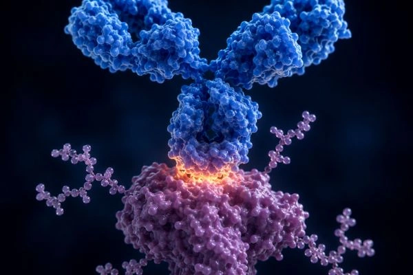
Most HCG immunoassays use a sandwich, or two-site, format. A capture antibody is immobilized on a solid surface such as a microplate well or a bead, the sample is introduced so the analyte binds the capture antibody, and a labeled detection antibody then binds a second epitope on the same molecule. The result is a capture-analyte-detection complex that generates a measurable signal proportional to the amount of HCG present.
Antibody pair selection drives the performance of this design. Targeting unique epitopes on the beta subunit improves specificity, while some assays pair an anti-alpha capture antibody with an anti-beta detection antibody. The two-site format suits HCG well because the molecule is large enough to present spatially separated epitopes that two antibodies can bind simultaneously. Researchers should treat antibody pairing as a deliberate design decision rather than a default, since it defines both sensitivity and the molecular forms the assay recognizes.
Common Immunoassay Formats
Several immunoassay formats are available for HCG detection, each with a distinct balance of sensitivity, throughput, and instrumentation.
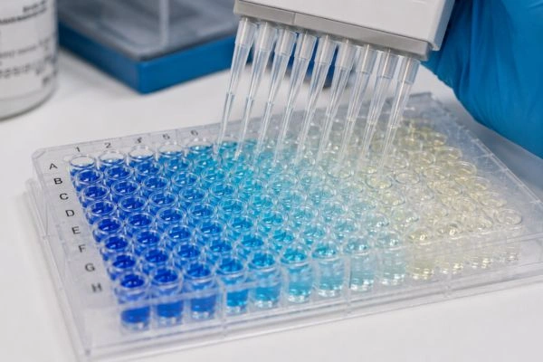
Enzyme-linked immunosorbent assay (ELISA) is the workhorse plate-based format. It uses enzyme labels such as horseradish peroxidase or alkaline phosphatase together with a chromogenic substrate like TMB, producing a color change read as absorbance on a plate reader. ELISA offers cost-effective, scalable throughput and remains a standard starting point for many laboratories.
Chemiluminescent immunoassay (CLIA) and electrochemiluminescence (ECL) use luminescent labels such as acridinium esters or ruthenium complexes and measure emitted light. These formats deliver high sensitivity and a wide dynamic range, and they are common on automated analytical platforms.
Fluorescence immunoassay, including time-resolved fluorescence (TRFIA), uses fluorophore or lanthanide chelate labels such as europium. Time-resolved detection separates the measurement from short-lived background fluorescence, which improves sensitivity and signal clarity.
Radioimmunoassay (RIA) is a foundational format that uses a radiolabeled tracer, historically iodine-125, in a competitive design. While it established many of the principles still used today, it has largely been superseded by non-radioactive formats because of the handling, licensing, and disposal demands of radioactivity. It remains relevant for historical and methodological context.
Lateral flow immunoassay (LFIA) is a rapid membrane-based format that uses colloidal gold or colored latex labels to produce a visible qualitative or semi-quantitative readout. It is useful for fast screening in research settings where speed matters more than precise quantification.
Detection Labels and Signal Generation
The label is what converts an antibody binding event into a measurable signal, so it shapes the analytical character of the entire assay. Enzymes generate amplified colorimetric or luminescent output, chemiluminescent and electrochemiluminescent labels emit light directly, fluorophores and lanthanide chelates fluoresce under defined excitation, and colloidal gold produces a visible colored line. Label choice governs the assay’s sensitivity, dynamic range, and instrumentation requirements. Researchers select the label by matching it to the analytical goal, weighing the need for sensitivity against available equipment and the desired throughput.
Antibody Specificity and HCG Variant Recognition
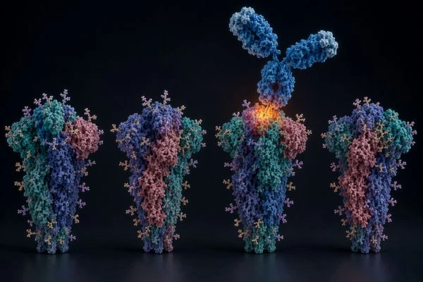
HCG research samples can contain multiple molecular forms, including intact HCG, free beta subunit, hyperglycosylated HCG, nicked forms, and the beta core fragment. Each form presents its epitopes differently, so the antibody pair defines which species an assay actually detects. This makes specificity a central analytical consideration rather than a technical footnote.
A key specificity challenge is cross-reactivity with related glycoprotein hormones, since the alpha subunit is shared with luteinizing hormone, follicle-stimulating hormone, and thyroid-stimulating hormone. Antibodies directed at unique beta subunit epitopes minimize this cross-reactivity and improve selectivity for HCG. Researchers characterizing HCG samples should document which molecular forms their chosen antibodies recognize, because two assays can report different values on the same sample purely because of differing form recognition. Recording antibody clones and their epitope targets is therefore essential to interpreting results correctly.
Analytical Challenges to Control
Several phenomena can compromise HCG detection if they are not actively managed. The high-dose hook effect is one of the most important: at very high analyte concentrations, a sandwich assay can paradoxically produce a lower signal, leading to underreporting. Running sample dilutions guards against this distortion and confirms that a result sits within the validated range.
Interferences present a second challenge. Heterophilic antibodies and matrix components can generate false signals, so appropriate blocking reagents, controls, and sample handling reduce this risk. Standardization is a third consideration. Because HCG is inherently heterogeneous, calibrating against recognized reference preparations supports comparability across assays, instruments, and laboratories. Addressing these factors directly is what separates a robust HCG detection method from one that produces inconsistent data.
Beyond Immunoassay: Complementary Detection
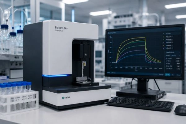
While immunoassays dominate HCG detection, complementary methods add specificity and structural detail. Liquid chromatography coupled to tandem mass spectrometry (LC-MS/MS) can distinguish HCG isoforms by mass and sequence, offering orthogonal confirmation that antibody-based methods alone cannot provide. Biosensor approaches, including surface plasmon resonance (SPR) and electrochemical immunosensors, measure binding in real time and support label-free or rapid detection. Bead-based multiplex immunoassays allow HCG to be measured alongside other analytes in a single run. Pairing an immunoassay with one of these orthogonal methods strengthens confidence in detection results and helps resolve ambiguous findings.
Validating a Reliable Detection Workflow
To establish dependable HCG detection, validate the method against defined analytical parameters. Determine the limit of detection and the limit of quantification, confirm linearity across the intended working range, and measure precision through intra-assay and inter-assay coefficients of variation. Assess accuracy through recovery experiments, and verify specificity against related glycoproteins and known HCG variants.
Documentation is equally important. Recording reagents, antibody clones, calibrators, and data processing settings ensures the method is reproducible and that results remain comparable over time. A validated, well-documented detection workflow is the foundation of credible HCG research data, and it allows other researchers to reproduce and verify the work.
Conclusion
Immunoassays remain the primary tools for detecting HCG in research samples, with sandwich formats, ELISA, chemiluminescence, fluorescence, and lateral flow each offering a distinct profile of sensitivity and throughput. Detection accuracy ultimately rests on antibody specificity, careful control of interferences and the hook effect, and consistent calibration against reference materials. By pairing a validated immunoassay with orthogonal methods such as mass spectrometry or biosensors where needed, analytical chemists and academic researchers can characterize HCG with the rigor and reproducibility their work demands.
Disclaimer: All products and reference materials referenced are intended strictly for laboratory research and educational use only. HCG research samples are not for human consumption, are not for diagnostic or therapeutic use, and are not intended to treat, cure, or prevent any condition. Nothing in this article constitutes medical, clinical, or professional advice. This content is provided for informational and educational purposes for qualified researchers and academic professionals. Handling, storage, and use of research materials must comply with all applicable institutional guidelines, safety protocols, and local, state, and federal regulations.
Frequently Asked Questions
Which immunoassay format works best for HCG detection?
Select the format that fits your analytical goal. ELISA delivers cost-effective, scalable plate-based throughput and works well as a starting point. Choose chemiluminescent (CLIA) or electrochemiluminescent (ECL) formats when you need high sensitivity and a wide dynamic range on automated platforms. Apply time-resolved fluorescence to separate the measurement from short-lived background and sharpen signal clarity. Reach for lateral flow when speed matters more than precise quantification. Match the format to your sensitivity, throughput, and instrumentation requirements rather than defaulting to a single option.
How does a sandwich immunoassay detect HCG?
A capture antibody immobilized on a microplate well or bead binds the HCG analyte, then a labeled detection antibody binds a second epitope on the same molecule. This forms a capture-analyte-detection complex that produces a signal proportional to the amount of HCG present. Select your antibody pair deliberately, since targeting unique beta subunit epitopes improves specificity and the two antibodies must bind spatially separated epitopes at the same time. Treat antibody pairing as a core design decision because it defines both sensitivity and the molecular forms your assay recognizes.
What is the high-dose hook effect and how do I prevent it?
The high-dose hook effect occurs when very high analyte concentrations cause a sandwich assay to paradoxically produce a lower signal, which leads to underreporting. Guard against it by running serial sample dilutions and confirming that your result falls within the validated range. Dilute any sample you suspect contains a high HCG concentration, then compare the diluted and undiluted readings. Build dilution checks into your workflow to prevent this distortion and keep your quantification accurate.
Why does antibody specificity matter for HCG detection?
HCG research samples can contain intact HCG, free beta subunit, hyperglycosylated HCG, nicked forms, and the beta core fragment, and each form presents its epitopes differently. Your antibody pair determines which species the assay actually detects. Specificity also controls cross-reactivity, because the shared alpha subunit can trigger signals from luteinizing hormone, follicle-stimulating hormone, and thyroid-stimulating hormone. Direct your antibodies at unique beta subunit epitopes to minimize cross-reactivity, and record antibody clones and their epitope targets so you can interpret and reproduce your results correctly.
Should I add complementary methods to my HCG immunoassay?
Add orthogonal methods whenever you need structural detail or want to resolve ambiguous findings. Use LC-MS/MS to distinguish HCG isoforms by mass and sequence, supplying confirmation that antibody-based methods alone cannot provide. Apply surface plasmon resonance or electrochemical immunosensors for real-time, label-free binding measurements. Run bead-based multiplex immunoassays to measure HCG alongside other analytes in a single experiment. Pairing your immunoassay with one of these methods strengthens confidence in your detection results and supports the reproducibility your research demands.